Review
. 2019 Nov 27:13:528.
doi: 10.3389/fncel.2019.00528. eCollection 2019.

Traumatic Brain Injuries: Pathophysiology and Potential Therapeutic Targets

Affiliations
Review

Traumatic Brain Injuries: Pathophysiology and Potential Therapeutic Targets

Si Yun Ng et al. Front Cell Neurosci. .

Abstract

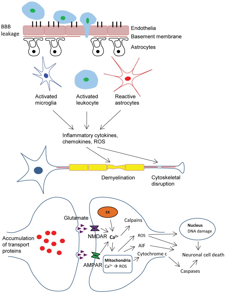
Traumatic brain injury (TBI) remains one of the leading causes of morbidity and mortality amongst civilians and military personnel globally. Despite advances in our knowledge of the complex pathophysiology of TBI, the underlying mechanisms are yet to be fully elucidated. While initial brain insult involves acute and irreversible primary damage to the parenchyma, the ensuing secondary brain injuries often progress slowly over months to years, hence providing a window for therapeutic interventions. To date, hallmark events during delayed secondary CNS damage include Wallerian degeneration of axons, mitochondrial dysfunction, excitotoxicity, oxidative stress and apoptotic cell death of neurons and glia. Extensive research has been directed to the identification of druggable targets associated with these processes. Furthermore, tremendous effort has been put forth to improve the bioavailability of therapeutics to CNS by devising strategies for efficient, specific and controlled delivery of bioactive agents to cellular targets. Here, we give an overview of the pathophysiology of TBI and the underlying molecular mechanisms, followed by an update on novel therapeutic targets and agents. Recent development of various approaches of drug delivery to the CNS is also discussed.

Keywords: CNS trauma; biopolymers; cell penetrating proteins; controlled drug release; neuronal regeneration; secondary injuries.


Figures

Figure 1
Figure 1
Schematic representation of pathophysiology of traumatic brain injury (TBI). BBB dysfunction caused by TBI insult allows transmigration of activated leukocytes into the injured brain parenchyma, which is facilitated by an upregulation of cell adhesion molecules. Activated leukocytes, microglia and astrocytes produce ROS and inflammatory molecules such as cytokines and chemokines that contribute to demyelination and disruption of axonal cytoskeleton, leading to axonal swelling and accumulation of transport proteins at the terminals, hence compromising neuronal activity. Progressive axonal damage results in neurodegeneration. In addition, astrogliosis at the lesion site causes glial scar formation, which creates a non-permissive environment that impedes axonal regeneration. On the other hand, excessive accumulation of glutamate and aspartate neurotransmitters in the synaptic space due to spillage from severed neurons, glutamate-induced aggravated release from pre-synaptic nerve terminals and impaired reuptake mechanisms in traumatic and ischemic brain activate NMDA and AMDA receptors located on post-synaptic membranes, which allow the influx of calcium ions. Together with the release of Ca2+ ions from intracellular store (ER), these events lead to the production of ROS and activation of calpains. As a result of mitochondrial dysfunction, molecules such as apoptosis-inducing factor (AIF) and cytochrome c are released into the cytosol. These cellular and molecular events including the interaction of Fas-Fas ligand ultimately lead to caspase-dependent and -independent neuronal cell death. BBB, blood-brain-barrier; ROS, reactive oxygen species; AMPA, α-amino-3-hydroxy-5-methyl-4-isoxazolepropionic acid; NMDA, N-methyl-d-aspartate; ER, endoplasmic reticulum.

References

    1. Ahn M. J., Sherwood E. R., Prough D. S., Lin C. Y., Dewitt D. S. (2004). The effects of traumatic brain injury on cerebral blood flow and brain tissue nitric oxide levels and cytokine expression. J. Neurotrauma 10, 1431–1442. 10.1089/neu.2004.21.1431 - DOI - PubMed
    1. Ai J., Liu E., Wang J., Chen Y., Yu J., Baker A. J. (2007). Calpain inhibitor MDL-28170 reduces the functional and structural deterioration of corpus callosum following fluid percussion injury. J. Neurotrauma 24, 960–978. 10.1089/neu.2006.0224 - DOI - PubMed
    1. Aktories K., Wilde C., Vogelsgesang M. (2005). Rho-modifying C3-like ADP-ribosyltransferases. Rev. Physiol. Biochem. Pharmacol. 152, 1–22. 10.1007/s10254-004-0034-4 - DOI - PubMed
    1. Alessandri B., Rice A. C., Levasseur J., Deford M., Hamm R. J., Bullock M. R. (2002). Cyclosporin A improves brain tissue oxygen consumption and learning/memory performance after lateral fluid percussion injury in rats. J. Neurotrauma 19, 829–841. 10.1089/08977150260190429 - DOI - PubMed
    1. Alvarez-Erviti L., Seow Y., Yin H., Betts C., Lakhal S., Wood M. J. (2011). Delivery of siRNA to the mouse brain by systemic injection of targeted exosomes. Nat. Biotechnol. 29, 341–345. 10.1038/nbt.1807 - DOI - PubMed