. 2019 Nov 11:2019:7548160.
doi: 10.1155/2019/7548160. eCollection 2019.

A Three-Dimensional Culture Model of Reversibly Quiescent Myogenic Cells

Affiliations

A Three-Dimensional Culture Model of Reversibly Quiescent Myogenic Cells

Salvatore Aguanno et al. Stem Cells Int. .

Abstract

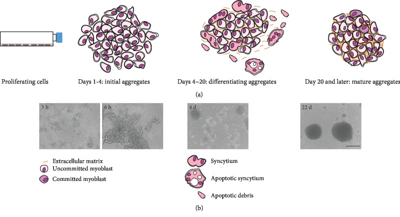
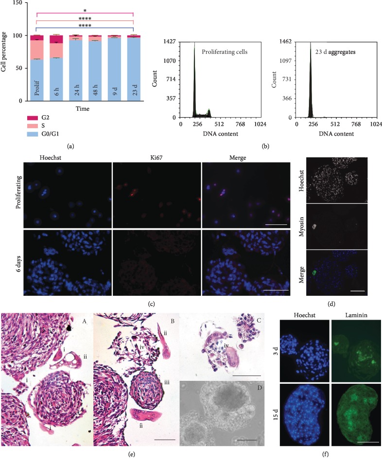
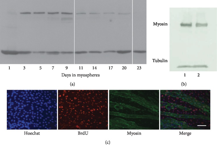
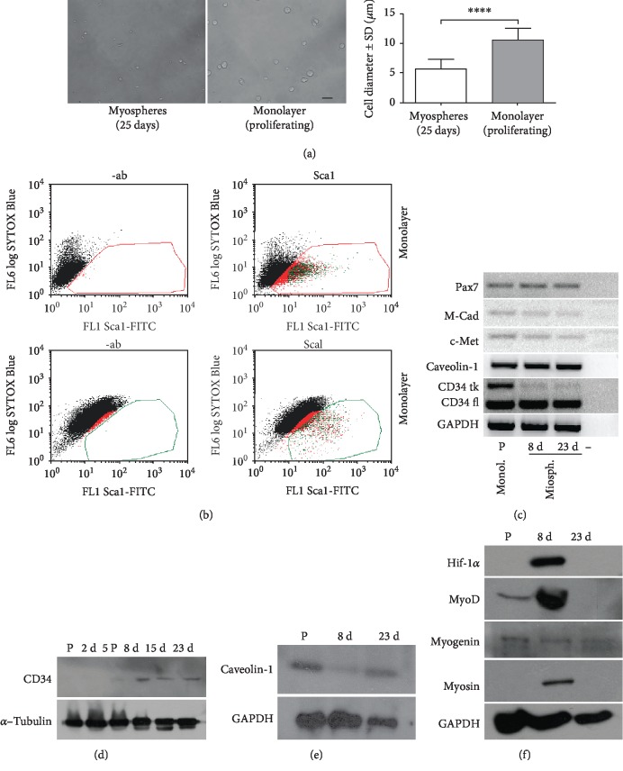
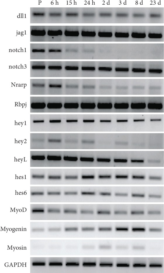
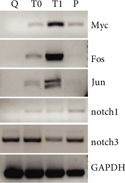
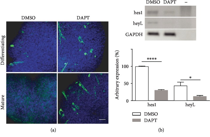
Satellite cells (SC) are the stem cells of skeletal muscles. They are quiescent in adult animals but resume proliferation to allow muscle hypertrophy or regeneration after injury. The mechanisms balancing quiescence, self-renewal, and differentiation of SC are difficult to analyze in vivo owing to their complexity and in vitro because the staminal character of SC is lost when they are removed from the niche and is not adequately reproduced in the culture models currently available. To overcome these difficulties, we set up a culture model of the myogenic C2C12 cell line in suspension. When C2C12 cells are cultured in suspension, they enter a state of quiescence and form three-dimensional aggregates (myospheres) that produce the extracellular matrix and express markers of quiescent SC. In the initial phase of culture, a portion of the cells fuses in syncytia and abandons the myospheres. The remaining cells are mononucleated and quiescent but resume proliferation and differentiation when plated in a monolayer. The notch pathway controls the quiescent state of the cells as shown by the fact that its inhibition leads to the resumption of differentiation. Within this context, notch3 appears to play a central role in the activity of this pathway since the expression of notch1 declines soon after aggregation. In summary, the culture model of C2C12 in suspension may be used to study the cellular interactions of muscle stem cells and the pathways controlling SC quiescence entrance and maintenance.


Conflict of interest statement

The authors declare no competing or financial interests.

Figures

Figure 1
Figure 1
Scheme of myosphere evolution shown according to (a) culture time and (b) contrast phase microscopy.
Figure 2
Figure 2
Characterization of C2C12 myospheres. (a) Bar chart from cytofluorometer data showing the proportion of cells (±SD) in different phases of the cycle, in a monolayer and in myospheres of different ages. 0.01 < p < 0.05 and ∗∗∗∗p < 0.0001. (b) Cytofluorometer plot comparing the cell cycle in cells proliferating in a monolayer and in 23-day myospheres. (c) Ki67 expression in a monolayer and 6-day myospheres. (d) Myosin expression evaluated by immunofluorescence. A labelled syncytium is at the periphery of a 9-day myosphere. (e) H&E staining of 9-day myospheres containing differentiating cells. Syncytia leaving the myospheres or internally located are indicated with (i), syncytia isolated in the medium after exit from a myosphere with (ii). (iii) indicates a mature myosphere that does not appear to contain syncytia. (e) in B and C, the symbol (iv) labels small myospheres dissolving after the detachment of syncytia. (f) Accumulation of extracellular matrix proteins in myospheres over time. Laminin expression in 3-day and 12-day myospheres. Scale bar: 50 μm in (c), (d), and (f) and 100 μm in (e).
Figure 3
Figure 3
Trend of myogenic differentiation during myosphere maturation and differentiation potential retained in quiescent cells of mature myospheres. (a) Time course of myosin expression in myospheres by western blot from the 1st to 23rd day of culture. (b) The cells of mature myospheres retain the capacity to proliferate and differentiate. The western blot quantitatively compares myosin expression in a monolayer: (1) original C2C12 cells that were never grown as myospheres and (2) cells derived from the dissociation of 23-day myospheres plated in a monolayer and allowed to differentiate. (c) C2C12 cells dissociated from 23-day myospheres (mature); replated in a monolayer, the cells incorporate BrdU while proliferating before differentiating and expressing myosin. Scale bar: 50 μm.
Figure 4
Figure 4
Cell size and marker expression. (a) Representative image comparing C2C12 cells shortly after dissociation of 25-day myospheres and detachment from the monolayer. The graph on the side shows the range of cell diameters. ∗∗∗∗p < 0.0001. (b) Flow cytometric profile of Sca1 expression in cells from myospheres (25 days) and from a monolayer. (c) Semiquantitative RT-PCR profile of common quiescent satellite cell markers in differentiating and mature myospheres. Myogenin and myosin indicate the presence of differentiating cells. P: proliferating cells in monolayer. (d) Time course of CD34 expression in myospheres by western blot. (e) Caveolin-1 expression in proliferating cells and in differentiating and mature myospheres by western blot. (f) Western blot comparing hif-1α expression in proliferating cells and myospheres, with the expression of myogenic markers.
Figure 5
Figure 5
Expression of notch pathway genes in proliferating cells and in myospheres at different stages by RT-PCR. The figure summarizes experiments performed more than three times.
Figure 6
Figure 6
Dissociation of myospheres induces changes in the transcription pattern of the cells. Q: RNA extraction performed from mature myospheres; T0: cells processed for RNA extraction after myosphere dissociation was completed; T1: cells maintained in suspension in the culture medium at 37°C for 1 h after myosphere dissociation; P: cells from a monolayer.
Figure 7
Figure 7
Differentiation resumes in mature myospheres following inhibition of the notch pathway. (a) Confocal image of whole-mount myosin immunofluorescence on differentiating (9 days) and mature (25 days) myospheres treated for 5 days with DAPT and respective DMSO controls. The effect of notch pathway inhibition is not particularly evident in differentiating myospheres because syncytia continue to leave the myospheres as they are formed. By contrast, DAPT resumes differentiation in mature myospheres, which retain the syncytia. Scale bar: 50 μm (b) RT-PCR showing the change of hes1 and heyL expression in mature myospheres in response to 5 μM DAPT treatment. Below the densitometric analysis of hes1 and heyL bands. 0.01 < p < 0.05 and ∗∗∗∗p < 0.0001.

References

    1. Charge S. B., Rudnicki M. A. Cellular and molecular regulation of muscle regeneration. Physiological Reviews. 2004;84(1):209–238. doi: 10.1152/physrev.00019.2003. - DOI - PubMed
    1. Coller H. A., Sang L., Roberts J. M. A new description of cellular quiescence. PLoS Biology. 2006;4(3, article e83) doi: 10.1371/journal.pbio.0040083. - DOI - PMC - PubMed
    1. Liu L., Cheung T. H., Charville G. W., et al. Chromatin modifications as determinants of muscle stem cell quiescence and chronological aging. Cell Reports. 2013;4(1):189–204. doi: 10.1016/j.celrep.2013.05.043. - DOI - PMC - PubMed
    1. Dhawan J., Rando T. A. Stem cells in postnatal myogenesis: molecular mechanisms of satellite cell quiescence, activation and replenishment. Trends in Cell Biology. 2005;15(12):666–673. doi: 10.1016/j.tcb.2005.10.007. - DOI - PubMed
    1. Cosgrove B. D., Sacco A., Gilbert P. M., Blau H. M. A home away from home: challenges and opportunities in engineering in vitro muscle satellite cell niches. Differentiation. 2009;78(2-3):185–194. doi: 10.1016/j.diff.2009.08.004. - DOI - PMC - PubMed

LinkOut - more resources