. 2019 Nov 15:2019:7989276.
doi: 10.1155/2019/7989276. eCollection 2019.

Enhancement of Quercetin-Induced Apoptosis by Cotreatment with Autophagy Inhibitor Is Associated with Augmentation of BAK-Dependent Mitochondrial Pathway in Jurkat T Cells

Affiliations

Enhancement of Quercetin-Induced Apoptosis by Cotreatment with Autophagy Inhibitor Is Associated with Augmentation of BAK-Dependent Mitochondrial Pathway in Jurkat T Cells

Eun Ji Ha et al. Oxid Med Cell Longev. .

Abstract

A flavonoid antioxidant quercetin promotes dose-dependent activation of the ATM-CHK-p53 pathway, downregulation of antiapoptotic survivin, and upregulation of proapoptotic NOXA in human T cell acute lymphoblastic leukemia Jurkat clones (J/Neo and J/BCL-XL). However, the downregulation of antiapoptotic BAG3 and MCL-1 occurred in J/Neo cells but not in J/BCL-XL cells overexpressing BCL-XL. Additionally, several BCL-XL-sensitive intrinsic mitochondrial apoptotic events including apoptotic sub-G1 cell accumulation, TUNEL-positive DNA fragmentation, BAK activation, mitochondrial membrane potential (Δψm) loss, caspase-9/caspase-8/caspase-3 activation, and PARP cleavage were induced only in J/Neo cells. Both cytosolic and mitochondrial ROS levels were elevated in quercetin-treated J/Neo cells; however, the ROS elevations were almost completely abrogated in J/BCL-XL cells, suggesting the ROS elevations were downstream of BCL-XL-sensitive mitochondrial damage and dysfunction. Wild-type A3, FADD-deficient I2.1, and caspase-8-deficient I9.2 Jurkat clones exhibited similar susceptibilities to the cytotoxicity of quercetin, excluding an involvement of extrinsic pathway in triggering the apoptosis. The autophagic events such as attenuation of AKT-mTOR pathway, formation of acridine orange-stainable acidic vesicular organelles, conversion of microtubule-associated protein 1 light chain 3-I (LC3-I) to LC3-II, and downregulation of p62/SQSTM1 level were detected in quercetin-treated J/Neo and J/BCL-XL cells, regardless of BCL-XL overexpression. Cotreatment with the autophagy inhibitor (3-methyladenine, LY294002, or chloroquine) resulted in a significant enhancement of quercetin-induced BAK activation and subsequently the mitochondrial damage-mediated apoptosis pathway by augmenting the downregulation of BAG3 and MCL-1 levels in J/Neo cells. These results demonstrated that quercetin induces intrinsic apoptosis and cytoprotective autophagy, and autophagy inhibition can potentiate BAK-dependent apoptotic activity of quercetin in Jurkat T cells.


Conflict of interest statement

The authors declare no conflict of interest.

Figures
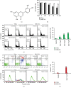
Figure 1
Figure 1
Cytotoxicity of quercetin toward Jurkat T cells was mainly exerted by the induction of BCL-XL-sensitive apoptosis without necrosis. (a) The chemical structure of quercetin. (b) For cell viability analysis, individual cells (5 × 10⁴/well) were incubated with vehicle (0.1% DMSO) or quercetin at indicated doses in a 96-well plate for 11 h and further incubated with MTT for 4 h to measure cell viability. Data are expressed as means ± SD (n = 3 with three replicates per independent experiment). (c, d) Cell cycle distribution was measured by flow cytometric analysis with PI staining. (e, f) Annexin V-positive apoptotic cells were determined by flow cytometric analysis with FITC-Annexin V/PI double staining. The forward scatter properties of unstained live, early apoptotic, and late apoptotic cells were measured to analyze alterations in cell size during the induced apoptosis. A representative study is shown and two additional experiments yielded similar results. All data in bar graphs represent the means of triplicate experiments. Error bars represent standard deviations with and ∗∗ indicating P < 0.05 and P < 0.01, respectively, compared with the control.
Figure 2
Figure 2
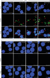
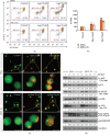
TUNEL-positive DNA fragmentation was observed in quercetin-treated J/Neo cells in a dose-dependent manner but not in J/BCL-XL cells. Individual cells were incubated at a density of 5 × 10⁵/ml with vehicle or indicated concentrations of quercetin for 11 h. Apoptotic DNA fragmentation analysis was performed using a TUNEL assay as described in Materials and Methods. Symbols: red arrowhead: TUNEL-positive DNA fragmentation. The scale bar represents a length of 10 μm in the images. A representative study is shown; two additional experiments yielded similar results.
Figure 3
Figure 3
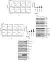
Quercetin induces mitochondria-dependent apoptotic events and DNA damage-mediated activation of the ATM-CHK-p53 pathway in Jurkat T cells. (a, b) BAK activation and (c, d) Δψm loss were determined by flow cytometry in J/Neo and J/BCL-XL cells treated with vehicle or quercetin at indicated doses for 11 h. (e, f) Total cell lysates from equivalent cultures were prepared, and western blot analyses of survivin, NOXA, BAK, BID, BIM, caspase-9, caspase-8, caspase-3, PARP, p-ATM (Ser-1981), ATM, p-CHK1 (Ser-317), CHK1, p-CHK2 (Ser-19), CHK2, p-p53 (Ser-15), p53, BAG3, BCL-XL, BCL-2, MCL-1, and GAPDH were carried out as described in Materials and Methods. A representative study is shown and two additional experiments yielded similar results. Error bars represent standard deviations with and ∗∗ indicating P < 0.05 and P < 0.01, respectively, compared with the control.
Figure 4
Figure 4
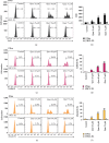
Cytosolic and mitochondrial ROS levels were commonly enhanced in quercetin-treated J/Neo cells but not in J/BCL-XL cells. (a, b) The intracellular ROS, (c, d) cytosolic ROS, and (e, f) mitochondrial ROS levels in J/Neo and J/BCL-XL cells treated with vehicle or quercetin at indicated doses for 11 h were analyzed using flow cytometry with DHE, CellROX Deep Red, and MitoSOX Red staining, respectively, and indicated by the MFI or percentage of the cells. A representative study is shown and two additional experiments yielded similar results. Error bars represent standard deviations with and ∗∗ indicating P < 0.05 and P < 0.01, respectively, compared with the control.
Figure 5
Figure 5
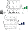
The sensitivity of FADD- and capase-8-positive wild-type Jurkat clone A3 to the apoptogenic activity of quercetin was similar to that of FADD-deficient Jurkat T cell clone I2.1 and caspase-8-deficient Jurkat T cell clone I9.2. (a) Exponentially growing individual cells were subjected to western blot analyses of caspase-8, FADD, and GAPDH as described in Materials and Methods. (b–e) After A3, I2.1, or I9.2 cells (5 × 10⁵ cells/ml) were incubated with vehicle or quercetin at indicated doses for 11 h, the cells were stained with PI and with DiOC6 for flow cytometric analysis of the cell cycle state and Δψm loss, respectively, as described in Materials and Methods. A representative study is shown and two additional experiments yielded similar results. Error bars represent standard deviations with ∗∗ indicating P < 0.01, compared with the control.
Figure 6
Figure 6
Quercetin induces autophagy in J/Neo and J/BCL-XL cells. (a–c) After cells were treated with vehicle or quercetin at indicated doses (25 and 50 μM) for 11 h, the enhancement of acridine orange- (AO-) Red fluorescence was analyzed by flow cytometry and fluorescence microscopy, respectively. The AO-Red fluorescence indicates the formation of AVOs and autolysosome vacuoles, resulting from autophagy induction, and the AO-Green fluorescence indicates AO staining of DNA/RNA in cells. Error bars represent standard deviations with and ∗∗ indicating P < 0.05 and P < 0.01, respectively, compared with the control. (d) Western blot analyses of p-AKT (Thr-308), AKT, p-mTOR (Ser-2448), mTOR, p-ULK (Ser-757), LC3-I/LC3-II, p62/SQSTM1, and GAPDH were performed as described in Materials and Methods. A representative study is shown and two additional experiments yielded similar results.
Figure 7
Figure 7
Cotreatment of J/Neo cells with pharmacological inhibitors (3-MA, LY, or CQ) of autophagy promotes quercetin-induced sub-G1 cell accumulation, Δψm loss, and BAK activation. (a–f) After treatment with 50 μM quercetin in the absence or presence of either 500 μM 3-MA, 20 μM LY, or 50 μM CQ for 7 h, the cells were harvested and subjected to flow cytometry to analyze the percentage of apoptotic sub-G1 cells, Δψm loss, and BAK activation. A representative study is shown and two additional experiments yielded similar results. Error bars represent standard deviations with and ∗∗ indicating P < 0.05 and P < 0.01, respectively, compared with the control.
Figure 8
Figure 8
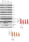
Cotreatment of J/Neo cells with pharmacological inhibitors (3-MA, LY, or CQ) of autophagy promotes quercetin-induced downregulation of BAG3 and MCL-1 levels, caspase-9/caspase-8 activation, and PARP cleavage. (a) After treatment with 50 μM quercetin in the absence or presence of either 500 μM 3-MA, 20 μM LY, or 50 μM CQ for 7 h, the cells were harvested and subjected to western blot analyses of BAG3, MCL-1, survivin, NOXA, BAK, caspase-9, caspase-8, PARP, and GAPDH as described in Materials and Methods. A representative study is shown and two additional experiments yielded similar results. (b, c) The arbitrary densitometric units of BAG3 and MCL-1 were normalized to those of GAPDH. Error bars represent standard deviations with and ∗∗ indicating P < 0.05 and P < 0.01, respectively, compared with the control.

References

    1. Aifantis I., Raetz E., Buonamici S. Molecular pathogenesis of T-cell leukaemia and lymphoma. Nature Reviews Immunology. 2008;8(5):380–390. doi: 10.1038/nri2304. - DOI - PubMed
    1. Van Vlierberghe P., Ferrando A. The molecular basis of T cell acute lymphoblastic leukemia. Journal of Clinical Investigation. 2012;122(10):3398–3406. doi: 10.1172/jci61269. - DOI - PMC - PubMed
    1. Hunger S. P., Lu X., Devidas M., et al. Improved survival for children and adolescents with acute lymphoblastic leukemia between 1990 and 2005: a report from the children’s oncology group. Journal of Clinical Oncology. 2012;30(14):1663–1669. doi: 10.1200/jco.2011.37.8018. - DOI - PMC - PubMed
    1. Amaravadi R. K., Thompson C. B. The roles of therapy-induced autophagy and necrosis in cancer treatment. Clinical Cancer Research. 2007;13(24):7271–7279. doi: 10.1158/1078-0432.ccr-07-1595. - DOI - PubMed
    1. Mansilla S., Llovera L., Portugal J. Chemotherapeutic targeting of cell death pathways. Anti-Cancer Agents in Medicinal Chemistry. 2012;12(3):226–238. doi: 10.2174/187152012800228805. - DOI - PubMed

MeSH terms