Review
. 2019 Dec 2:9:97.
doi: 10.1186/s13578-019-0361-4. eCollection 2019.

Signaling pathways involved in colorectal cancer progression

Affiliations
Review

Signaling pathways involved in colorectal cancer progression

Zahra Koveitypour et al. Cell Biosci. .

Abstract

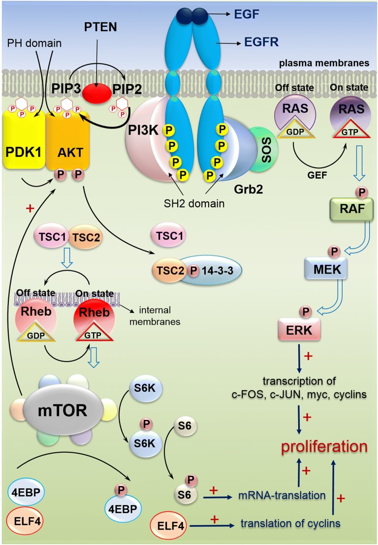
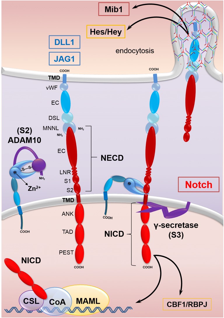
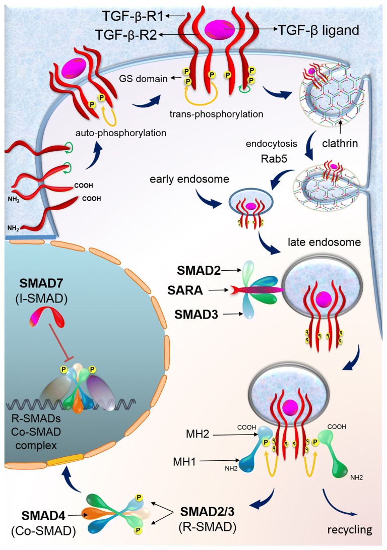
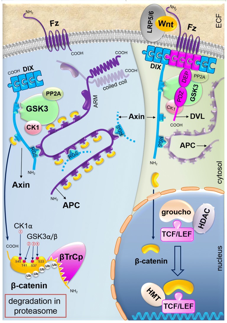
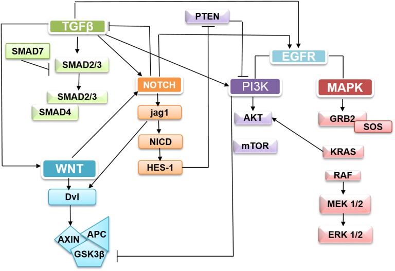
Colorectal cancer (CRC) is the fourth leading cause of the worldwide cancer mortality. Different molecular mechanisms have been attributed to the development and progress of CRC. In this review, we will focus on the mitogen-activated protein kinase (MAPK) cascades downstream of the epidermal growth factor receptor (EGFR), Notch, PI3K/AKT pathway, transforming growth factor-β (TGF-β), and Wnt signaling pathways. Various mutations in the components of these signaling pathways have been linked to the development of CRC. Accordingly, numerous efforts have been carried out to target the signaling pathways to develop novel therapeutic approaches. Herein, we review the signaling pathways involved in the incidence and progression of CRC, and the strategies for the therapy targeting components of signaling pathways in CRC.

Keywords: Colorectal cancer; EGFR; MAPK; Notch; TGF-β.


Conflict of interest statement

Competing interestsThe authors declare that they have no competing interests.

Figures

Fig. 1
Fig. 1
EGFR and PI3K signaling pathways in CRC. The binding of EGF to the extracellular domain of EGFR, induces dimerization, and activation of intrinsic kinase activity. The proteins, those are recruited to active EGFR include a number of Src homology 2 (SH2) proteins. One of the adaptor proteins, GRB2 recruits SOS to the membrane. SOS activates GDP/GTP exchange which recruits RAF to the membrane. RAF phosphorylates MEKs, which then activates the extracellular signal regulated kinase (ERK). Phosphorylated ERK translocates to nucleus and activates transcription factors leading to expression of the target genes such as c-FOS, c-JUN and myc [4]. GRB2 recruits PI3Ks, another major mediator of EGFR signaling pathway. PI3Ks converts PIP2 to PIP3. PIP3 binds to PH domain of AKT and recruits it to plasma membrane. PDK1 phosphorylates AKT which in turn regulates the activity of various proteins that mediate cell survival. Activated AKT inhibits TSC2 via phosphorylation. Inactive TSC1/2 is unable to bind RAS homolog enriched in brain (RHEB), which subsequently enables its activation of mTORC1 at the surface of lysosome. Upon activation, mTORC1 regulates many cellular functions, such as cell growth, protein synthesis and autophagy via S6 kinase (S6K; RPS6K) and eukaryotic translation initiation factor 4E-binding protein 1 (4E-BP1; EIF4EBP1) [68]
Fig. 2
Fig. 2
Notch signaling pathways in CRC. Mind bomb-1 (Mib1), an E3 ubiquitin ligase, promotes the endocytosis of Notch ligands. The extracellular domain of Notch ligands (DLL1 or JAG1 shown) consists of an N-terminal MNNL domain, DSL domain, EC and vWF [69]. The Notch receptors are transmembrane proteins containing extracellular or NECD (EC, LNR, S1 and S2), transmembrane domain or TMD and intracellular domains or NICD (ANK, TAD and PEST) [19]. Activation of Notch pathway is initiated by binding of Notch ligands to Notch receptors which leads to their conformational changes. Then, S2 cleavage site is exposed for ADAM to remove the extracellular region. Subsequently, S3 cleavage occurs by γ-secretase which removes the transmembrane region and releases NICD. Then NICD translocates into the nucleus, and binds to the inactive CSL transcription factor which forms a complex [20]. Instead co-activators, such as MAML could bind to CSL, resulting in activating of this transcriptional complex [21]. Finally, expression of target genes, including Hes family, is induced [18]
Fig. 3
Fig. 3
TGF-β signaling pathways in CRC. TGF-β receptors internalization occurs through clathrin-dependent or lipid-raft-dependent pathways. However, clathrin-dependent endocytosis of TGF-β receptors positively facilitates TGF-β signaling while internalization through lipid raft/caveolae exerts an inhibitory effect [70]. The internalized receptors are targeted to distinct destination through different functions of Rab5 GTPases. Binding of TGF-β ligands to TGFBR2 triggers initiation of TGF-β signaling. By binding TGF-β to TGFBR2, TGFBR2 recruits and phosphorylates TGFBR1, stimulating the protein kinase activity of TGFBR1, in which TGFBR1 is activated. Then, R-SMAD proteins or SMAD2 and SMAD3 are phosphorylated and activated by the activated TGFBR1, thereby allowing them to bind to SMAD4. As a result, the R-SMAD effectors make a complex with SMAD4 and SMAD. This complex migrates to the nucleus to regulate transcription of the target genes [28, 29]. In addition, SMAD7 has an inhibitory effect on the interaction of R-SMAD with TGFBR1 [30]. Multiple proteins contribute to the recruitment of R-SMAD proteins to the TGFBR1s and enhance SMAD activation, such as SARA [28]
Fig. 4
Fig. 4
WNT signaling pathways in CRC. Accumulation of secretory Wnt ligands leads to the interaction between FZD and LRP, resulting in the activation of the DVL protein [38]. The DVL is activated and phosphorylated and translocated to the FZD receptor. The β-catenin dissociates from the degradation complex and accumulates in the cytoplasm followed by migration to the nucleus [36]. β-catenin, which is accumulated in the nucleus, could be coupled with TCF or LEF, thereby triggers activating of the expression of target genes involved in pathophysiology of CRC. These target genes are involved in the proliferation and transmission. In the absence of Wnt induction, the cytoplasmic β-catenin exists in the destruction complex, and it is phosphorylated by CK and GSK3β. Subsequently this complex recruits β-TrCP E3 linker and then degrades β-catenin via the proteasome [4]
Fig. 5
Fig. 5
The Notch, PI3K, TGF-β, Wnt/B-catenin, and EGFR/Ras signaling pathways are interacting with each other. Notch signaling regulates Wnt pathway through disheveled and Wnt signaling actives Jagged-1 [56]. Also Notch stimulates EGFR and suppresses the activity of TGF-β pathway [20, 57]. HES1 inhibits PTEN thus activates PI3/AKT pathway in CRC [20]. PI3K can inactivates GSK3β as their downstream target and KRAS can actives PI3K [58]. TGF-β superfamily signaling can activates oncogenic pathways (PI3K/AKT, MAPK/ERK, WNT, and Notch) in CRC [31]

References

    1. Wong A, Ma BB. Personalizing therapy for colorectal cancer. Clin Gastroenterol Hepatol. 2014;12(1):139–144. doi: 10.1016/j.cgh.2013.08.040. - DOI - PubMed
    1. Fearon ER. Molecular genetics of colorectal cancer. Annu Rev Pathol. 2011;6:479–507. doi: 10.1146/annurev-pathol-011110-130235. - DOI - PubMed
    1. Tabana YM, Dahham SS, Shah AM, Majid A. Major signaling pathways of colorectal carcinogenesis. Recent Adv Colon Cancer. 2016;1:1–2. doi: 10.14302/issn.2471-7061.jcrc-14-579. - DOI
    1. Jeong WJ, Ro EJ, Choi KY. Interaction between Wnt/β-catenin and RAS-ERK pathways and an anti-cancer strategy via degradations of β-catenin and RAS by targeting the Wnt/β-catenin pathway. NPJ Precis Oncol. 2018;2(1):5. doi: 10.1038/s41698-018-0049-y. - DOI - PMC - PubMed
    1. Wu WK, Wang XJ, Cheng AS, Luo MX, Ng SS, To KF, et al. Dysregulation and crosstalk of cellular signaling pathways in colon carcinogenesis. Crit Rev Oncol Hematol. 2013;86(3):251–277. doi: 10.1016/j.critrevonc.2012.11.009. - DOI - PubMed