

Optical Coherence Tomography Angiography Assessed Retinal and Choroidal Microvasculature Features in Patients with Retinitis Pigmentosa: A Meta-Analysis
- PMID: 31828118
- PMCID: PMC6881583
- DOI: 10.1155/2019/6723917
Optical Coherence Tomography Angiography Assessed Retinal and Choroidal Microvasculature Features in Patients with Retinitis Pigmentosa: A Meta-Analysis
Abstract
Background: The aim of this study was to assess the retinal and choroidal microvasculature features using optical coherence tomography angiography (OCTA) in patients with retinitis pigmentosa (RP).
Methods: This study was a meta-analysis of relevant published studies that were included after a comprehensive search of PubMed, Embase, Cochrane Library, and Web of Science databases. Mean difference (MD) with a 95% confidence interval was used to assess continuous variable outcomes. Heterogeneity was evaluated using the chi-squared test based on the values of P and I 2.
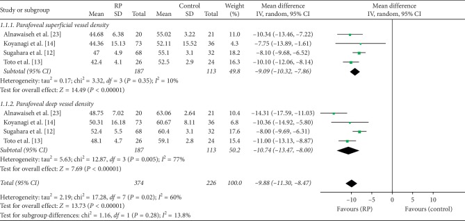
Results: Seven studies were included in this meta-analysis. The vessel density values measured in the superficial and deep foveal zones of RP patients using OCTA were significantly lower than the recorded values in the control groups (MD = -3.58, P=0.04; MD = -4.93, P=0.02, respectively). The superficial and deep parafoveal vessel density values measured with OCTA were also significantly lower in RP patients than in control groups (MD = -9.09, P < 0.00001; MD = -10.74, P < 0.00001, respectively); for choriocapillaris vessel density, there was no statistically significant difference between RP patients and controls (MD = -1.33, P=0.09). The deep foveal avascular zone (FAZ) was significantly larger in RP patients than in controls (MD = 0.15, P=0.01), whereas there was no significant difference in the superficial foveal avascular zones in the two groups (MD = 0.08, P=0.11).
Conclusions: We showed that retinal and choroidal vessels were attenuated in RP patients. Additionally, we revealed that the FAZ was larger in RP patients, especially the deep FAZ. OCTA may become a useful modality in the diagnosis and monitoring of patients with RP.
Copyright © 2019 Ling Ling et al.
Conflict of interest statement
The authors have no conflicts of interest to this submission.
Figures


References
-
- Berson E. L. Retinitis pigmentosa. The friedenwald lecture. Investigative Ophthalmology & Visual Science. 1993;34(5):1659–1676. - PubMed
Publication types
MeSH terms
LinkOut - more resources
Full Text Sources
