. 2020 Feb;42(1):169-182.
doi: 10.1007/s11357-019-00133-8. Epub 2019 Dec 11.

Lumbar spinal cord microglia exhibited increased activation in aging dogs compared with young adult dogs

Affiliations

Lumbar spinal cord microglia exhibited increased activation in aging dogs compared with young adult dogs

Christine M Toedebusch et al. Geroscience. 2020 Feb.

Abstract

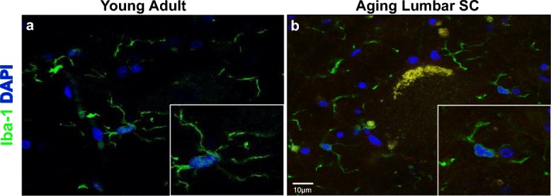
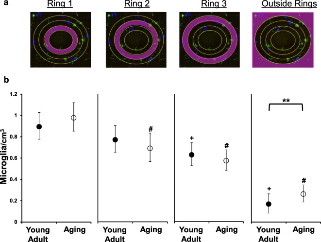
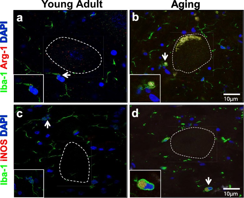
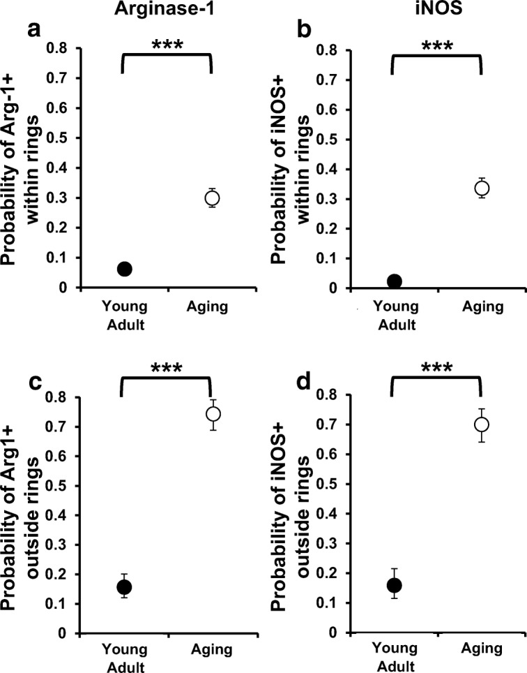
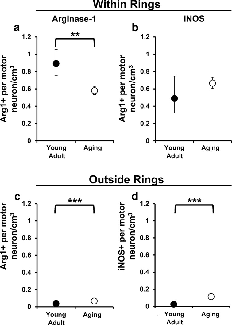
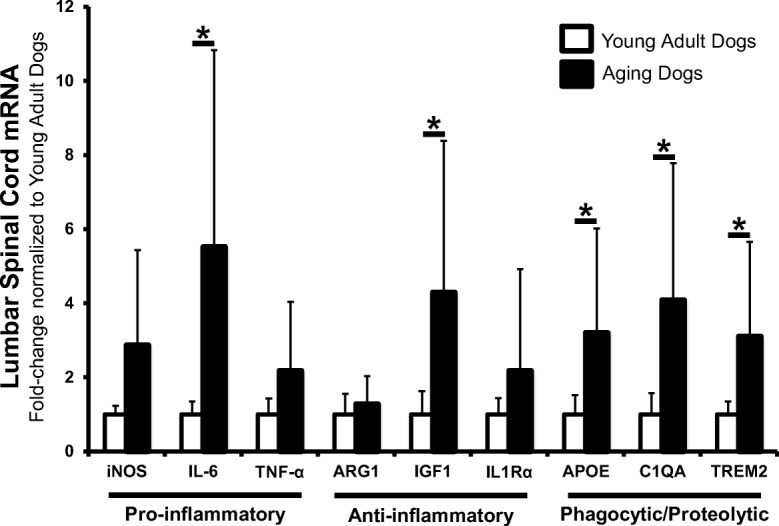
Altered microglia function contributes to loss of CNS homeostasis during aging in the brain. Few studies have evaluated age-related alterations in spinal cord microglia. We previously demonstrated that lumbar spinal cord microglial expression of inducible nitric oxide synthase (iNOS) was equivalent between aging, neurologically normal dogs and dogs with canine degenerative myelopathy (Toedebusch et al. 2018, Mol Cell Neurosci. 88, 148-157). This unexpected finding suggested that microglia in aging spinal cord have a pro-inflammatory polarization. In this study, we reexamined our microglial results (Toedebusch et al. 2018, Mol Cell Neurosci. 88, 148-157) within the context of aging rather than disease by comparing microglia in aging versus young adult dogs. For both aging and young adult dogs, the density of microglia was significantly higher closest to the motor neuron cell body. However, there was no difference in densities between aging versus young adult dogs at all distances except for the furthest distance analyzed. The number of motor neurons with polarized microglia was higher in aging dogs; yet, the density per motor neuron of arginase-1-expressing microglia was reduced in aging dogs compared with young adult dogs. Finally, aging dogs had increased steady-state mRNA levels for genes consistent with activated microglia compared with young adult dogs. However, altered mRNA levels were limited to the lumbar spinal cord. These data suggested that aging dog spinal cord microglia exhibit regional immunophenotypic differences, which may render lumbar motor neurons more susceptible to age-related pathological insults.

Keywords: Aging; Canine; Microglia; Neurodegeneration.


Figures

Fig. 1
Fig. 1
Microglia in aging lumbar spinal cords exhibited morphology consistent with activation. Representative images of lumbar spinal cord microglia (Iba-1; green). a Microglia from young adult dogs tended to have long, branching processes. b A subset of microglia from aging dogs exhibited decreased ramification of their processes, which was consistent with activation
Fig. 2
Fig. 2
Microglial density was greatest in close approximation to motor neurons in both young adult and aging dogs. a Using a modified Sholl’s method (Sholl 1953), concentric rings were placed around each motor neuron cell body. Each ring increased in diameter by 6 μM, the average microglial cell body diameter (originally published in Toedebusch et al. 2018). The total numbers of microglia cells within each ring, and outside the ring system, were quantified. b A generalized linear mixed effect model with nested effects identified an interaction between ring and group. When the number of microglia within each ring for young adult dogs was compared, there was no difference between microglial number between ring 1 and ring 2 (223 observations; p = 0.1723), but ring 1 had increased microglia versus ring 3 (p < 0.0001) and versus outside the rings (p < 0.0001). In aging dogs, ring 1 had significantly increased number of microglia from all other rings and outside the rings (227 observations; p < 0.0001 all comparisons). Comparison of microglial number between young adult and aging dogs did not reveal differences in any of the rings (ring 1, p = 0.5567; ring 2, p = 0.4763; ring 3, p = 0.5554). However, the average number of microglia was increased in aging dogs compared with young adult dogs outside the ring system (p = 0.0025). Data is displayed as the estimated mean with 95% confidence interval. **, difference between young adult and aging dogs, p < 0.01; +, difference from ring 1 young adult dogs, p < 0.001; #, difference from ring 1 aging dogs, p < 0.001
Fig. 3
Fig. 3
Aging, but not young adult, microglia expressed both arginase-1 and inducible nitric oxide synthase (iNOS). Representative images of lumbar spinal cord microglia (Iba-1; green) in close proximity to motor neurons (outlined in white dotted line). a When observed, positive immunoreactivity for arginase-1 (Arg-1; orange) was punctate near the microglia nucleus in young adult dogs. b Diffuse arginase-1 immunoreactivity was commonly observed in the microglial cell body in aging dogs (originally published in Toedebusch et al. 2018). c The majority of young adult microglia did not demonstrate immunoreactivity for iNOS. d iNOS immunoreactivity localizes to the microglia cell body in aging dogs (originally published in Toedebusch et al. 2018). Scale bar 10 μM
Fig. 4
Fig. 4
The number of polarized microglia near motor neurons was increased in aging dogs. There was a greater probability of identifying microglia cells that expressed arginase-1 or iNOS close to motor neurons (a, b) or outside the ring system (c, d) in aging dogs relative to young adult dogs (p < 0.001 for all comparisons). Moreover, in both groups, this probability was higher outside the ring system (young adult dogs P = 0.0011; aging dogs p < 0.001). ***, difference between young adult and aging dogs, p < 0.001
Fig. 5
Fig. 5
Density of arginase-1-expressing microglial near motor neurons was decreased in aging dogs. We examined the number of arginase-1- (Arg-1+) or iNOS-expressing (iNOS+) microglia per motor neuron to determine the average density of each phenotype within (a, b) and outside (c, d) the ring system. a Compared with young adult dogs, Arg-1+ microglia per motor neuron was decreased in aging dogs (p = 0.0027). b There was no difference in the number of iNOS-expressing microglial cells per motor neuron between groups (p = 0.3638). c Outside the ring system, Arg-1+ (c) and iNOS+ (d) microglial cells were increased in aging dogs compared with young adult dogs (for both comparisons p < 0.001). **, difference between young adult and aging dogs, p < 0.01; ***, difference between young adult and aging dogs, p < 0.001
Fig. 6
Fig. 6
Levels of microglia phenotypic markers were not different in the cervical spinal cord between young adult and aging dogs. mRNA levels of common pro-inflammatory (iNOS, IL-6, TNF-α), anti-inflammatory (Arg-1, IGF-1, IL1Rα), and phagocytic/proteolytic genes (ApoE, C1QA, TREM2) were not different between groups in hemisected C8 spinal cord segments. Data are displayed relative to young adult dogs. Young adult dogs, n = 6; aging dogs, n = 5. Comparisons based on unpaired t tests; bars represent group mean and standard error of the mean (SEM)
Fig. 7
Fig. 7
Aging dog lumbar spinal cord had increased levels of genes associated with reactive microglia. mRNA levels of all pro-inflammatory markers were increased in aging dogs. However, only increases measured in IL-6 reached statistical significance. mRNA levels of anti-inflammatory markers IGF1 and IL1Rα were increased with IL1Rα increases reaching statistical significance. All phagocytic/proteolytic mRNA levels (ApoE, C1QA, TREM2) were significantly increased in L4 spinal cord segments in aging dogs. Data are displayed relative to young adult dogs. Young adult dogs, n = 6; aging dogs, n = 5. *p < 0.05. Comparisons based on unpaired t tests; bars represent group mean and standard error of the mean (SEM).

References

    1. Ahn JH, Choi JH, Kim JS, Lee HJ, Lee CH, Yoo KY, Hwang IK, Lee YL, Shin HC, Won MH. Comparison of immunoreactivities in 4-HNE and superoxide dismutases in the cervical and the lumbar spinal cord between adult and aged dogs. Exp Gerontol. 2011;46:703–708. - PubMed
    1. Averill DR., Jr Degenerative myelopathy in the aging German Shepherd dog: clinical and pathologic findings. J Am Vet Med Assoc. 1973;15:1045–1051. - PubMed
    1. Awano T, Johnson GS, Wade CM, Katz ML, Johnson GC, Taylor JF, Perloski M, Biagi T, Baranowska I, Long S, March PA, Olby NJ, Shelton GD, Khan S, O'Brien DP, Lindblad-Toh K, Coates JR. Genome-wide association analysis reveals a SOD1 mutation in canine degenerative myelopathy that resembles amyotrophic lateral sclerosis. Proc Natl Acad Sci U S A. 2009;106:2794–2799. - PMC - PubMed
    1. Beers DR, Henkel JS, Xiao Q, Zhao W, Wang J, Yen AA, Siklos L, McKercher S, Appel SH. Wild-type microglia extend survival in PU.1 knockout mice with familial amyotrophic lateral sclerosis. Proc Natl Acad Sci U S A. 2006;103:16021–16026. - PMC - PubMed
    1. Bickford PC, et al. Aging leads to altered microglial function that reduces brain resiliency increasing vulnerability to neurodegenerative diseases. Exp Gerontol. 2017;94:4–8. - PMC - PubMed

Publication types