. 2020 Feb 3;17(2):392-403.
doi: 10.1021/acs.molpharmaceut.9b00589. Epub 2019 Dec 27.

Extracellular-Matrix-Anchored Click Motifs for Specific Tissue Targeting

Affiliations

Extracellular-Matrix-Anchored Click Motifs for Specific Tissue Targeting

Mary R Adams et al. Mol Pharm. .

Abstract

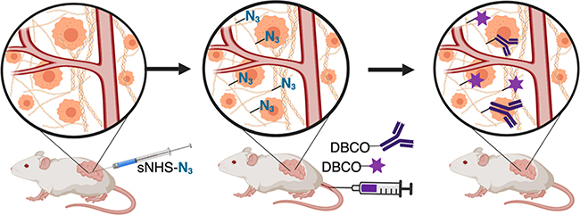
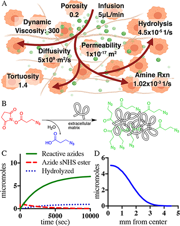
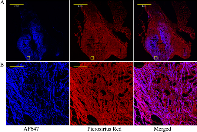
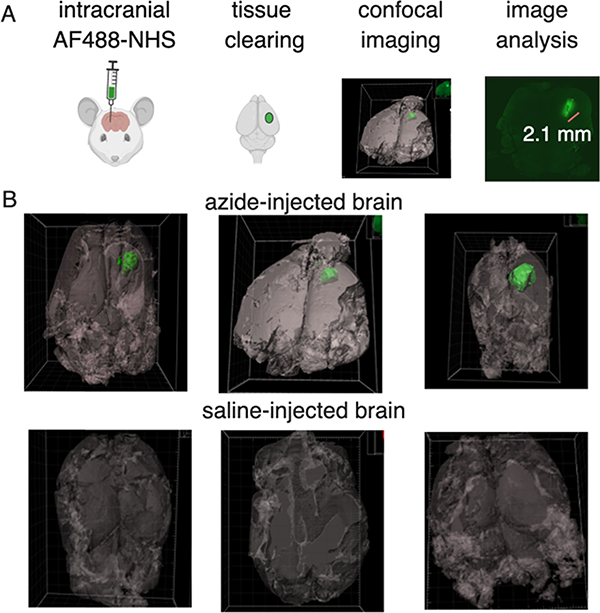
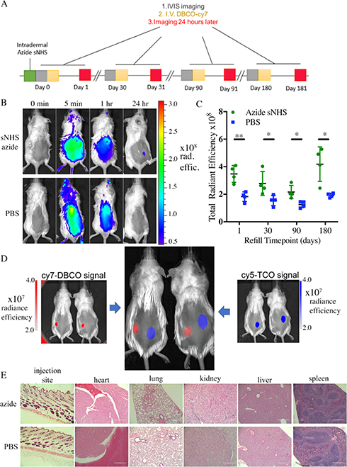
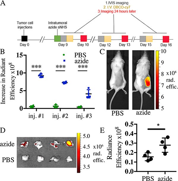
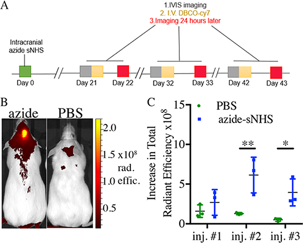
Local presentation of cancer drugs by injectable drug-eluting depots reduces systemic side effects and improves efficacy. However, local depots deplete their drug stores and are difficult to introduce into stiff tissues, or organs, such as the brain, that cannot accommodate increased pressure. We present a method for introducing targetable depots through injection of activated ester molecules into target tissues that react with and anchor themselves to the local extracellular matrix (ECM) and subsequently capture systemically administered small molecules through bioorthogonal click chemistry. A computational model of tissue-anchoring depot formation and distribution was verified by histological analysis and confocal imaging of cleared tissues. ECM-anchored click groups do not elicit any noticeable local or systemic toxicity or immune response and specifically capture systemically circulating molecules at intradermal, intratumoral, and intracranial sites for multiple months. Taken together, ECM anchoring of click chemistry motifs is a promising approach to specific targeting of both small and large therapeutics, enabling repeated local presentation for cancer therapy and other diseases.

Keywords: brain delivery; click chemistry; computer modeling; drug delivery; extracellular matrix; pancreatic cancer.


Conflict of interest statement

The authors declare no competing financial interest.

Figures

Figure 1.
Figure 1.
ECM anchoring to introduce click motifs in target tissues. Left to middle: injections of activated NHS esters anchor azide molecules to tissue ECM. Middle to right: intravascular administration of cyclooctyne conjugated to small molecules or antibodies allows selective capture and display at tumor sites.
Figure 2.
Figure 2.
Modeling of azide anchoring to tumor ECM with intratumoral fluid flow. (A) Schematic diagram of NHS-ester injection, aminolysis, and hydrolysis as well as COMSOL Multiphysics model parameters. (B) 0D model estimating the change in the concentration of the injected azide-sNHS ester, hydrolyzed species, and ECM-anchored azides over time. The expected reaction kinetics is further layered on a three-dimensional (3D) space-dependent model that leads to the results in (C). (C) Number of anchored azides available to bind to systemic DBCO molecules over mm from the center of the infusion needle in the tumor.
Figure 3.
Figure 3.
Imaging NHS-ester and extracellular matrix co-localization within a pancreatic tumor. (A) Whole tumor and (B) zoom-in images of the boxed area of fluorophore-NHS-injected tumors stained for extracellular proteins with picrosirius red. Pancreatic KPC 4662 tumors were injected intratumorally with AF647 NHS ester. After 24 h, tumors were excised, fixed, sectioned, and stained with picrosirius red. The scale bars for (A) and (B) are 2 mm and 100 μm, respectively.
Figure 4.
Figure 4.
Imaging NHS-ester distribution within a murine brain using model fluorescent NHS ester and tissue clearing. (A) Schematic of the azide depot visualization method from NHS injection, clearing, imaging, and analysis. (B) Representative green isosurfaces of the three Alexa Fluor 488 injected tumors and PBS-injected controls. Autofluorescence in tissues was visualized and set as a gray background.
Figure 5.
Figure 5.
Azide-sNHS ester depots allow long-term and repeated targeting with no apparent immunogenicity and are mutually compatible with tetrazine-TCO targeting for spatial separation of different regiments. (A) Timeline of systemic targeting of intradermal depots over long term. (B) Mice received an intradermal injection of azide-sNHS (50 μL of 0.2 M) or PBS and were administered iv DBCO-Cy7 and IVIS imaged before the dose and after 5 min, 1 h and 24 h. (C) Quantitation of systemic targeting of intradermal depots 1, 30, 90, and 180 days after intradermal injection of azide-sNHS (0.2 M, 50 μL) or PBS control. (D) 50 μL of methyltetrazine sNHS (right, 0.05 M) or azide-sNHS (left, 0.05 M) was injected intradermally on the dorsal flank of four mice. 1:1 DBCO-Cy7/TCO-Cy5 (200 μL) was injected iv (E) H&E staining of the skin injection site and major organs at 1 month for CD1 mice injected intradermally with 50 μL of azide-sNHS. Scale bar = 400 μm. Samples show mean ± standard error of the mean (SEM). *p < 0.05 by Student’s t-test. See Supporting Figures 7–10 for full IVIS images.
Figure 6.
Figure 6.
Click-specific capture of small molecules and antibodies at pancreatic tumor sites. (A) Timeline for the tumor-targeting experiment for (A)–(C). Increase in radiant efficiency over tumor ROI’s 24 h after iv DBCO-Cy7 administration. (C) Representative images of intratumoral azide-sNHS ester mice 24 h after iv dose of DBCO-Cy7 fluorophore. (D) Extracted azide-sNHS-infused tumors (top row) and PBS (bottom row) 24 h after iv dosing with DBCO- and Cy7-conjugated anti-PD1 antibody. (E) Quantification of radiant efficiency over tumor and underlying carcass ROI’s 24 h after iv DBCO-Cy7-antibody administration. Samples show mean ± SEM. *p < 0.05, **p < 0.01, ***p < 0.001 by Student’s t-test. See Supporting Figures 12–14 for full images.
Figure 7.
Figure 7.
Click-specific capture of small molecules in the brain. (A) Timeline of the intracranial-targeting experiment. (B) Representative images of azide-sNHS and control mice after three iv administrations of DBCO-Cy7. (C) Quantitation of intracranial ROI radiant efficiency measured 24 h after iv DBCO-Cy7 administration. Samples show mean ± SEM. *p < 0.05, **p < 0.01 by Student’s t-test. See Supporting Figures 19–21 for full images.

References

    1. Brudno Y; Pezone MJ; Snyder TK; Uzun O; Moody CT; Aizenberg M; Mooney DJ Replenishable Drug Depot to Combat Post-Resection Cancer Recurrence. Biomaterials 2018, 178, 373–382. - PMC - PubMed
    1. Mejia Oneto JM; Khan I; Seebald L; Royzen M In Vivo Bioorthogonal Chemistry Enables Local Hydrogel and Systemic Pro-Drug To Treat Soft Tissue Sarcoma. ACS Cent. Sci. 2016, 2, 476–482. - PMC - PubMed
    1. Ries F; Klastersky J Nephrotoxicity Induced by Cancer Chemotherapy With Special Emphasis on Cisplatin Toxicity. Am. J. Kidney Dis. 1986, 8, 368–379. - PubMed
    1. Segota E; Bukowski RM The Promise of Targeted Therapy: Cancer Drugs Become More Specific. Cleveland Clin. J. Med 2004, 71, 551–560. - PubMed
    1. Fakih M; Vincent M Adverse Events Associated with Anti-EGFR Therapies for the Treatment of Metastatic Colorectal Cancer. Curr. Oncol 2010, 17, S18–S30. - PMC - PubMed

Publication types

MeSH terms