. 2020 Jan 17;9(1):10-18.
doi: 10.1021/acssynbio.9b00347. Epub 2019 Dec 20.

Point-of-Use Detection of Environmental Fluoride via a Cell-Free Riboswitch-Based Biosensor

Affiliations

Point-of-Use Detection of Environmental Fluoride via a Cell-Free Riboswitch-Based Biosensor

Walter Thavarajah et al. ACS Synth Biol. .

Abstract

Advances in biosensor engineering have enabled the design of programmable molecular systems to detect a range of pathogens, nucleic acids, and chemicals. Here, we engineer and field-test a biosensor for fluoride, a major groundwater contaminant of global concern. The sensor consists of a cell-free system containing a DNA template that encodes a fluoride-responsive riboswitch regulating genes that produce a fluorescent or colorimetric output. Individual reactions can be lyophilized for long-term storage and detect fluoride at levels above 2 ppm, the Environmental Protection Agency's most stringent regulatory standard, in both laboratory and field conditions. Through onsite detection of fluoride in a real-world water source, this work provides a critical proof-of-principle for the future engineering of riboswitches and other biosensors to address challenges for global health and the environment.

Keywords: biosensor; cell-free systems; diagnostics; field use; riboswitches; water quality.


Figures

Figure 1.
Figure 1.. Cell-free fluoride biosensor engineering strategy.
(a) Schematic for lyophilization of a cell-free reaction in tubes or on paper disks. Rehydration with a water sample allows the designed biosensing reaction to proceed to yield a detectable signal. (b) Schematic for fluoride riboswitch-mediated transcriptional regulation in cell-free extract. The riboswitch folds cotranscriptionally into one of two mutually exclusive states, depending on the presence of fluoride. In the absence of fluoride, the riboswitch folds into a terminating hairpin, precluding downstream gene expression. Fluoride binding stabilizes a pseudoknot structure (red paired region, inset from PDB: 4ENC) that sequesters the terminator and enables the expression of downstream reporter genes. (c) Schematic of a cell-free fluoride biosensor, consisting of a DNA template encoding the fluoride riboswitch controlling the expression of sfGFP. Eight-hour endpoint fluorescence measurements for reactions containing NaF (dark green) or NaCl (gray) are shown below. Error bars represent one standard deviation from three technical replicates.
Figure 2.
Figure 2.. Riboswitch modularity allows fluorescent protein, RNA aptamer and enzymatic colorimetric reporter outputs.
Biosensor DNA template layouts and concentrations shown above reporter information and characterization data for that reporter. (a) Superfolder GFP (sfGFP) reporter (structure from PDB: 2B3P). Time course of fluorescence in the presence of 3.5 mM NaF (dark green), 0.2 mM NaF (light green), or 0 mM NaF (gray). (b) 3-way junction dimeric Broccoli reporter (structure predicted from NUPACK ). Time course of fluorescence in the presence of 3.5 mM NaF (dark green), 0.2 mM NaF (light green) and 0 mM NaF (gray). (c) Catechol (2,3)-dioxygenase (C23DO) reporter. Reaction scheme shows the cleavage of the colorless catechol molecule into the yellow 2-hydroxymuconate semialdehyde. Time course of absorbance at 385 nm in the presence of 3.5 mM NaF (orange), 0.2 mM NaF (yellow), and 0 mM NaF (gray). For each plot, trajectories represent average and error shading represents one standard deviation from three technical replicates. (a) and (b) are reported in mean equivalent fluorescence (MEF).
Figure 3.
Figure 3.. Colorimetric reporters enable fluoride sensing at environmentally relevant concentrations.
(a) Time course of 385 nm absorbance as measured by plate reader in the presence of 100 μM NaF (orange), 50 μM NaF (yellow), and 0 μM NaF (grey) using C23DO as a reporter and incubated at 30°C. Trajectories represent average and error shading represents one standard deviation from three technical replicates. (b) Color change observed after 1-hour for two different reporter template concentrations with and without 100 μM NaF. Tubes were mixed by pipetting and incubated at 37°C before image capture at 60 minutes. (c) Time lapse of rehydrated lyophilized reactions incubated at 37°C in the absence (top) and presence (bottom) of 1mM NaF.
Figure 4.
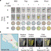
Figure 4.. The cell-free fluoride riboswitch biosensor functions with real-world water samples and is not impacted by long-term storage and distribution.
(a) Cell-free reactions rehydrated with various water samples with or without 1 mM NaF added. Lyophilized reactions in tubes are shown above lyophilized reactions on chromatography paper before and after one-hour incubation at 37°C. MQ = laboratory grade Milli-Q water; Tap = tap water; Lake = unfiltered Lake Michigan water; Pool = unfiltered outdoor pool water. Uncropped photos of chromatography paper experiments are available in Supplemental Figure 8. (b) Field testing of lyophilized cell-free reactions rehydrated with water sampled in Cartago, Costa Rica. Geographical map from OpenStreetMap. The positive control contained 1 mM NaF in the reaction before lyophilization. The negative control was rehydrated with Milli-Q water, and the positive control and each test were rehydrated with 20 μL of unprocessed field sample followed by body-heat incubation for five hours. Measured fluoride concentrations obtained using a fluoride sensing electrode. Field samples are from sites B and E in Supplementary Table 2.

References

    1. World Health Organization. World Health Statistics 2016: Monitoring Health for the SDGs Sustainable Development Goals; World Health Organization, 2016.
    1. Onda K; LoBuglio J; Bartram J. Global Access to Safe Water: Accounting for Water Quality and the Resulting Impact on MDG Progress. Int. J. Environ. Res. Public Health 2012, 9 (3), 880–894. - PMC - PubMed
    1. Maheshwari RC Fluoride in Drinking Water and Its Removal. J. Hazard. Mater 2006, 137 (1), 456–463. - PubMed
    1. World Health Organization. Guidelines for Drinking-Water Quality. WHO Chron. 2011, 38 (4), 104–108. - PubMed
    1. Zhou Y; Zhang JF; Yoon J. Fluorescence and Colorimetric Chemosensors for Fluoride-Ion Detection. Chem. Rev 2014, 114 (10), 5511–5571. - PubMed

Publication types