. 2020 Jan 17;15(1):93-102.
doi: 10.1021/acschembio.9b00639. Epub 2019 Dec 27.

Structural Basis for Finding OG Lesions and Avoiding Undamaged G by the DNA Glycosylase MutY

Affiliations

Structural Basis for Finding OG Lesions and Avoiding Undamaged G by the DNA Glycosylase MutY

L Peyton Russelburg et al. ACS Chem Biol. .

Abstract

The adenine glycosylase MutY selectively initiates repair of OG:A lesions and, by comparison, avoids G:A mispairs. The ability to distinguish these closely related substrates relies on the C-terminal domain of MutY, which structurally resembles MutT. To understand the mechanism for substrate specificity, we crystallized MutY in complex with DNA containing G across from the high-affinity azaribose transition state analogue. Our structure shows that G is accommodated by the OG site and highlights the role of a serine residue in OG versus G discrimination. The functional significance of Ser308 and its neighboring residues was evaluated by mutational analysis, revealing the critical importance of a β loop in the C-terminal domain for mutation suppression in cells, and biochemical performance in vitro. This loop comprising residues Phe307, Ser308, and His309 (Geobacillus stearothermophilus sequence positions) is conserved in MutY but absent in MutT and other DNA repair enzymes and may therefore serve as a MutY-specific target exploitable by chemical biological probes.


Figures

Figure 1.
Figure 1.
Overview of OG, TS and 1N analog structures. The chemical structure of guanine (G) and 8-oxo-7,8-dihydroguanine (OG) differ only slightly by changes in atoms attached at positions 7 and 8. The positively charged and extremely unstable oxacarbenium ion transition state is mimicked in terms of shape and charge by the inert 1N TS analog.
Figure 2.
Figure 2.
The GO repair pathway. MutY acts as a final line of defense to prevent G:C to T:A mutations that otherwise would accumulate consequent to reactive oxygen species (ROS).
Figure 3.
Figure 3.
Overview of Gs MutY in complex with DNA containing G paired with 1N. The N-terminal domain (cyan) and C-terminal domain (navy) surround the DNA (all atom stick model in the left-hand and expanded view; surface in the right-hand view). The 1N TS analog (black transparent surface) engages with catalytic residues of the active site found in the NTD. On the opposite DNA strand, the G base (green transparent surface) adopts the anti conformation stacked between a DNA base pair and Tyr88. FSH residues (purple) contact the DNA on the major groove side. The overall architecture of TSAC-G:1N shown here is highly comparable to previously determined structures of MutY in complex with DNA, especially the TSAC-OG:1N structure.
Figure 4.
Figure 4.
Difference maps define Ser308 side chain rotamer selection. Ser308 assumes different interactions and altered rotamer positions when G is present in the OG site compared to when OG is present in the OG site. (A) G (green) occupies the OG recognition site as seen in the TSAC-G:1N structure. (B) Ser308 (navy blue) in TSAC-G:1N showed a 5.0σ positive (green) peak and an 8.2σ negative (red) peak when modeled with the same rotamer as found in TSAC-OG:1N indicating incorrect model placement. (C) Ser308 of TSAC-OG:1N showed a 6.4σ positive peak and a 5.2σ negative peak when modeled with the same rotamer as found in TSAC-G:1N. (D) OG (yellow) as found in the TSAC-OG:1N structure. The 2|Fo|−|Fc| difference maps (gray) are contoured at 1.0σ. Positive and negative peaks in the |Fo|−|Fc| difference maps are contoured at 3.5σ.
Figure 5.
Figure 5.
Molecular interactions at the OG detection site. The OG site is made up of residues from both the NTD (cyan shading) and the CTD (violet shading). The G base interacts with rotamer “1” of Ser308 and OG interacts with rotamer “2”. For the G base X represents a lone-pair electron and Y represents a hydrogen atom. For the OG base X represents a hydrogen and Y represents an oxygen. Ser308 in the CTD is the only residue that makes interactions with OG-specific atoms, and these interactions are altered upon replacement of OG with G.
Figure 6.
Figure 6.
MutY alignment. The HXF(S/T)H region (shaded) is highly conserved among MutY from different species. Prepared with PROMALS3D.,
Figure 7.
Figure 7.. DNA-binding and kinetics.
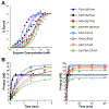
(A) DNA-binding isotherms measured by EMSA. EcNGsC MutY with FSH residues intact (FSH, blue triangles) and the S308A variant (FAH, black circles) each bound OG:FA-DNA (filled symbols) and G:FA-DNA (open symbols) with markedly different midpoints (see Table 2 for Kd values). The variants AAH (red squares) and Del FSH (green triangles) appear to all have very similar curves for the two substrates indicating a loss of OG versus G specificity. (B) Adenine glycosylase reactions. The left-hand panel highlights early timepoints and the right-hand panel shows full time-courses. EcNGsC MutY and the FAH variant processed OG:A and G:A with markedly different rates under single-turnover conditions at 60 °C (Table 3). Notably, this specificity indicator diminished or vanished for the variants with double-alanine replacement (AAH) and FSH residues deleted (Del FSH).
Scheme 1.
Scheme 1.
Minimal Kinetic Scheme for Glycosylase Reaction

References

    1. Lindahl T (1993) Instability and Decay of the Primary Structure of DNA. Nature 362, 709–715. - PubMed
    1. Steenken S, and Jovanovic SV (1997) How Easily Oxidizable Is DNA? One-Electron Reduction Potentials of Adenosine and Guanosine Radicals in Aqueous Solution. J. Am. Chem. Soc 119, 617–618.
    1. Michaels ML, and Miller JH (1992) The GO System Protects Organisms from the Mutagenic Effect of the Spontaneous Lesion 8-Hydroxyguanine (7,8-Dihydro-8-Oxoguanine). J. Bacteriol 174, 6321–6325. - PMC - PubMed
    1. Ito R, Hayakawa H, Sekiguchi M, and Ishibashi T (2005) Multiple Enzyme Activities of Escherichia Coli MutT Protein for Sanitization of DNA and RNA Precursor Pools. Biochemistry 44, 6670–6674. - PubMed
    1. Maki H, and Sekiguchi M (1992) MutT Protein Specifically Hydrolyses a Potent Mutagenic Substrate for DNA Synthesis. Nature 355, 273–275. - PubMed

Publication types