

Somatic mitochondrial mutation discovery using ultra-deep sequencing of the mitochondrial genome reveals spatial tumor heterogeneity in head and neck squamous cell carcinoma
- PMID: 31830557
- PMCID: PMC6980748
- DOI: 10.1016/j.canlet.2019.12.006
Somatic mitochondrial mutation discovery using ultra-deep sequencing of the mitochondrial genome reveals spatial tumor heterogeneity in head and neck squamous cell carcinoma
Abstract
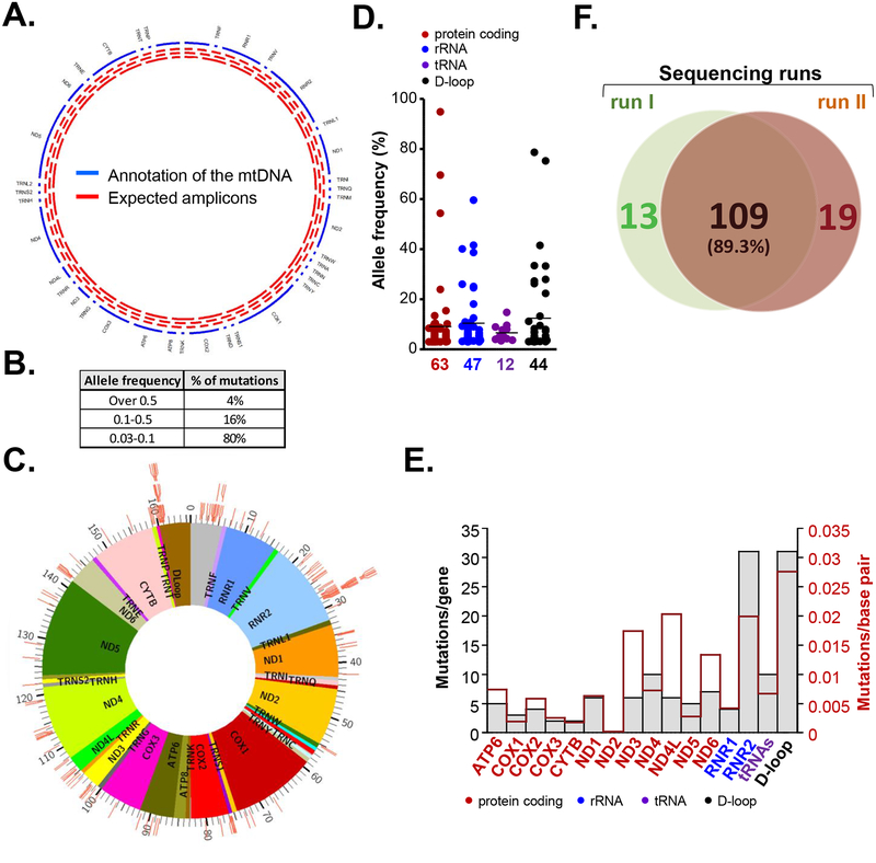
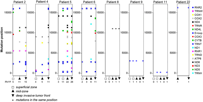
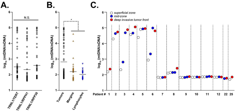
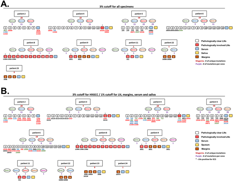
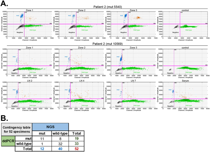
Mutations in mitochondrial DNA (mtDNA) have been linked to risk, progression, and treatment response of head and neck squamous cell carcinoma (HNSCC). Due to their clonal nature and high copy number, mitochondrial mutations could serve as powerful molecular markers for detection of cancer cells in bodily fluids, surgical margins, biopsies and lymph node (LN) metastasis, especially at sites where tumor involvement is not histologically apparent. Despite a pressing need for high-throughput, cost-effective mtDNA mutation profiling system, current methods for library preparation are still imperfect for detection of low prevalence heteroplasmic mutations. To this end, we have designed an ultra-deep amplicon-based sequencing library preparation approach that covers the entire mitochondrial genome. We sequenced mtDNA in 28 HNSCCs, matched LNs, surgical margins and bodily fluids, and applied multiregional sequencing approach on 14 primary tumors. Our results demonstrate that this quick, sensitive and cost-efficient method allows obtaining a snapshot on the mitochondrial heterogeneity, and can be used for detection of low frequency tumor-associated mtDNA mutations in LNs, sputum and serum specimens. These findings provide the foundation for using mitochondrial sequencing for risk assessment, early detection, and tumor surveillance.
Keywords: Cancer; Head and neck squamous cell carcinoma (HNSCC); Mitochondrial DNA (mtDNA); Mutations; Sequencing.
Copyright © 2019 Elsevier B.V. All rights reserved.
Conflict of interest statement
Figures
Similar articles
-
Mitochondrial DNA mutation in normal margins and tumors of recurrent head and neck squamous cell carcinoma patients.Cancer Prev Res (Phila). 2010 Sep;3(9):1205-11. doi: 10.1158/1940-6207.CAPR-10-0018. Epub 2010 Jul 26. Cancer Prev Res (Phila). 2010. PMID: 20660573 Free PMC article.
-
Mitochondrial DNA mutations in head and neck cancer are infrequent and lack prognostic utility.Br J Cancer. 2011 Apr 12;104(8):1319-24. doi: 10.1038/bjc.2011.96. Epub 2011 Mar 22. Br J Cancer. 2011. PMID: 21427725 Free PMC article.
-
Analysis of clinically relevant somatic mutations in high-risk head and neck cutaneous squamous cell carcinoma.Mod Pathol. 2018 Feb;31(2):275-287. doi: 10.1038/modpathol.2017.128. Epub 2017 Oct 6. Mod Pathol. 2018. PMID: 28984303
-
Association Between Mitochondrial DNA Copy Number and Head and Neck Squamous Cell Carcinoma: A Systematic Review and Dose-Response Meta-Analysis.Med Sci Monit. 2021 Jan 20;27:e928327. doi: 10.12659/MSM.928327. Med Sci Monit. 2021. PMID: 33468984 Free PMC article.
-
Mutations and polymorphisms in mitochondrial DNA in head and neck cancer cell lines.Acta Otorhinolaryngol Ital. 2006 Aug;26(4):185-90. Acta Otorhinolaryngol Ital. 2006. PMID: 18236634 Free PMC article. Review.
Cited by
-
Possible Immunotherapeutic Strategies Based on Carcinogen-Dependent Subgroup Classification for Oral Cancer.Front Mol Biosci. 2021 Aug 23;8:717038. doi: 10.3389/fmolb.2021.717038. eCollection 2021. Front Mol Biosci. 2021. PMID: 34497832 Free PMC article. Review.
-
Application of liquid biopsy as multi-functional biomarkers in head and neck cancer.Br J Cancer. 2022 Feb;126(3):361-370. doi: 10.1038/s41416-021-01626-0. Epub 2021 Dec 7. Br J Cancer. 2022. PMID: 34876674 Free PMC article. Review.
-
Characterization of ferroptosis driver gene signature in head and neck squamous cell carcinoma (HNSC).Am J Transl Res. 2023 Jul 15;15(7):4829-4850. eCollection 2023. Am J Transl Res. 2023. PMID: 37560204 Free PMC article.
-
Unexpected heterogeneity in oropharyngeal squamous cell tumors.Nat Genet. 2023 Apr;55(4):534-535. doi: 10.1038/s41588-023-01360-8. Nat Genet. 2023. PMID: 37016098 No abstract available.
-
Ultrasensitive detection of tumor-specific mutations in saliva of patients with oral cavity squamous cell carcinoma.Cancer. 2021 May 15;127(10):1576-1589. doi: 10.1002/cncr.33393. Epub 2020 Dec 21. Cancer. 2021. PMID: 33405231 Free PMC article.
References
-
- Kamangar F, Dores GM, Anderson WF, Patterns of cancer incidence, mortality, and prevalence across five continents: defining priorities to reduce cancer disparities in different geographic regions of the world, J Clin Oncol, 24 (2006) 2137–2150. - PubMed
-
- Jemal A, Siegel R, Xu J, Ward E, Cancer statistics, 2010, CA Cancer J Clin, 60 (2010) 277–300. - PubMed
-
- Parkin DM, Pisani P, Ferlay J, Global cancer statistics, CA Cancer J Clin, 49 (1999) 33–64, 31. - PubMed
-
- John Andrew Ridge RM, Miriam N Lango, Steven Feigenberg, Head and Neck Tumors, Haller Daniel G., Wagman Lawrence D., Camphausen Kevin A., Hoskins William J. (Eds) Cancer Management: A Multidisciplinary Approach., ed. 2013. (2013).
Publication types
MeSH terms
Substances
Grants and funding
LinkOut - more resources
Full Text Sources
Medical
