. 2020 Jan 15;26(2):477-486.
doi: 10.1158/1078-0432.CCR-19-2182. Epub 2019 Dec 12.

Ultrasound-mediated Delivery of Paclitaxel for Glioma: A Comparative Study of Distribution, Toxicity, and Efficacy of Albumin-bound Versus Cremophor Formulations

Affiliations

Ultrasound-mediated Delivery of Paclitaxel for Glioma: A Comparative Study of Distribution, Toxicity, and Efficacy of Albumin-bound Versus Cremophor Formulations

Daniel Y Zhang et al. Clin Cancer Res. .

Abstract

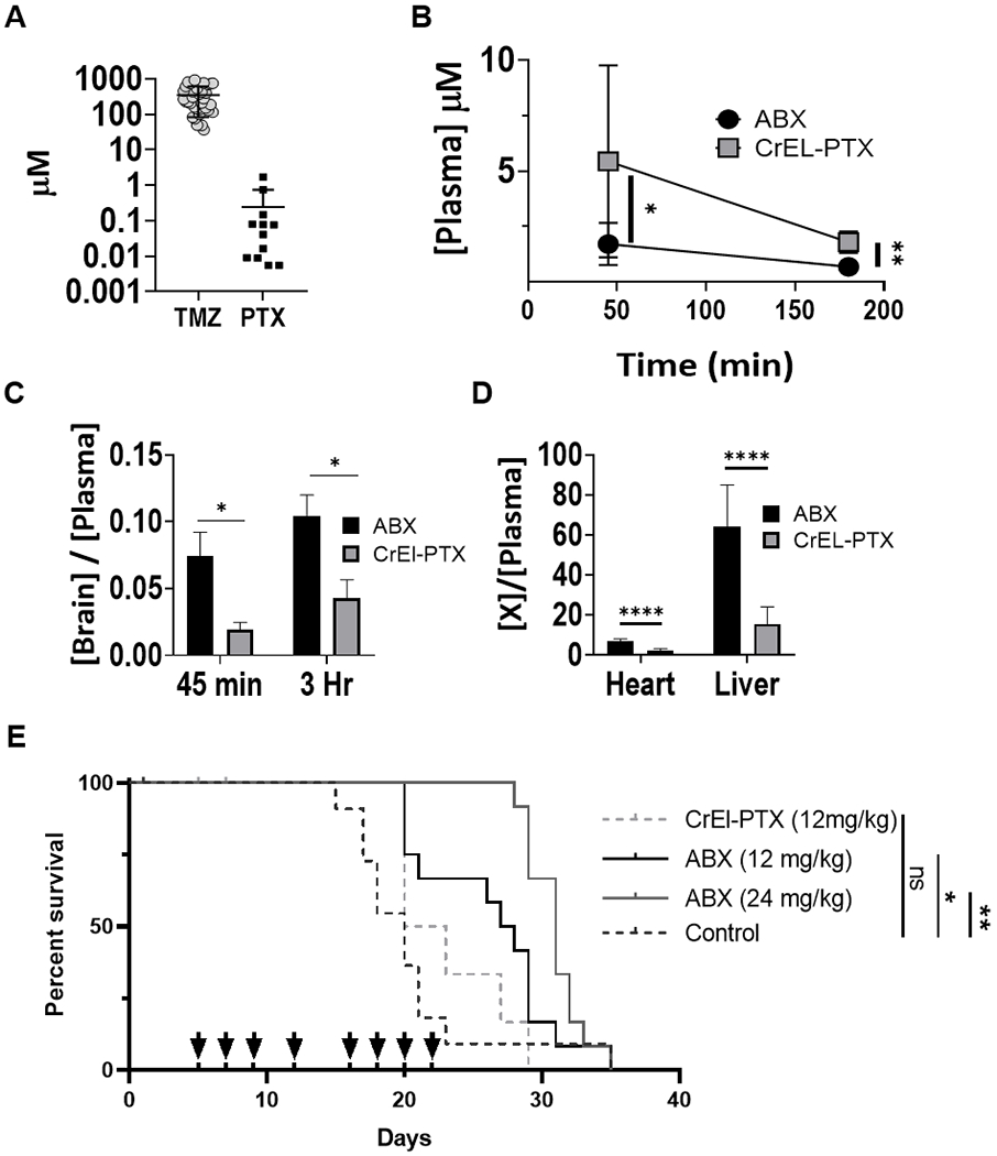
Purpose: Paclitaxel shows little benefit in the treatment of glioma due to poor penetration across the blood-brain barrier (BBB). Low-intensity pulsed ultrasound (LIPU) with microbubble injection transiently disrupts the BBB allowing for improved drug delivery to the brain. We investigated the distribution, toxicity, and efficacy of LIPU delivery of two different formulations of paclitaxel, albumin-bound paclitaxel (ABX) and paclitaxel dissolved in cremophor (CrEL-PTX), in preclinical glioma models.

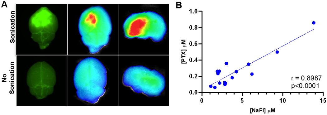
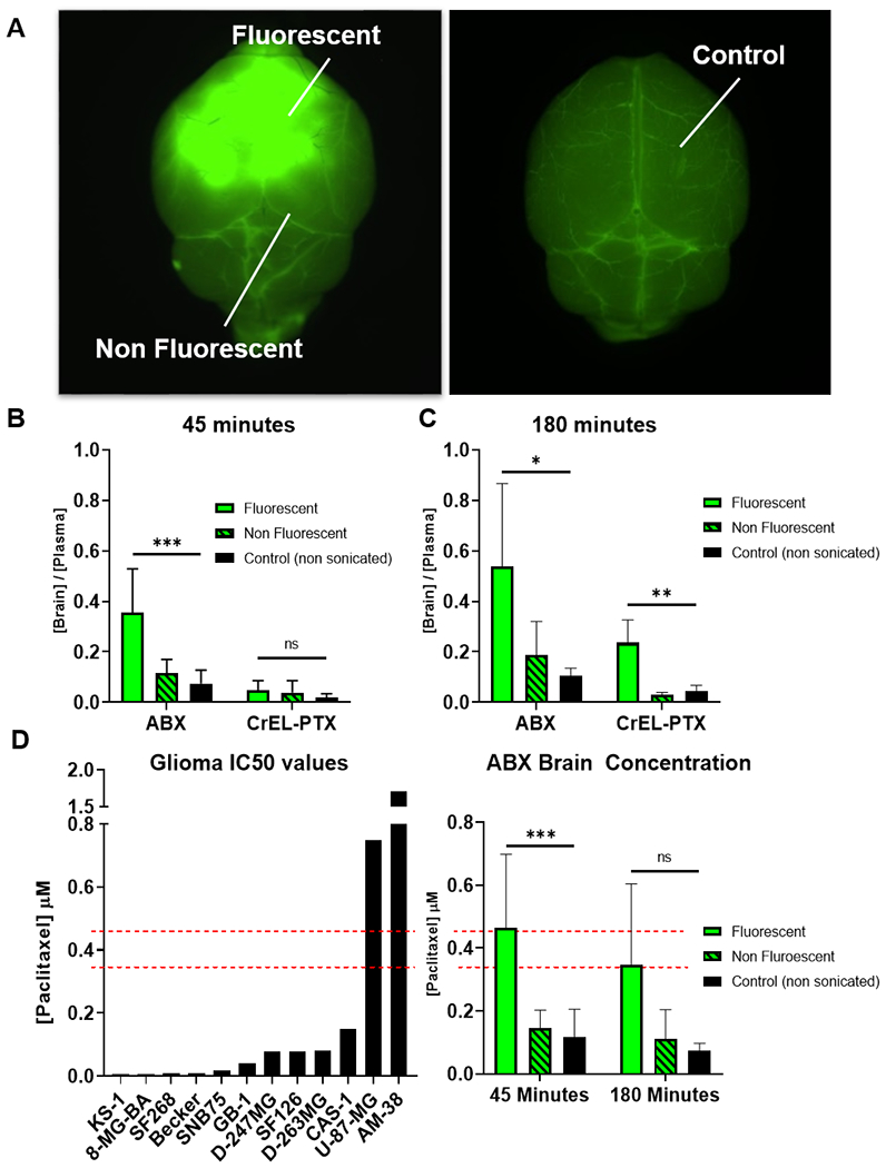
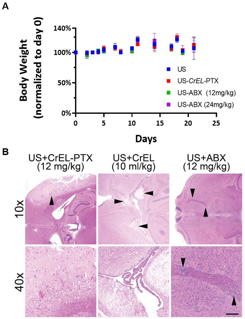
Experimental design: The efficacy and biodistribution of ABX and CrEL-PTX were compared with and without LIPU delivery. Antiglioma activity was evaluated in nude mice bearing intracranial patient-derived glioma xenografts (PDX). Paclitaxel biodistribution was determined in sonicated and nonsonicated nude mice. Sonications were performed using a 1 MHz LIPU device (SonoCloud), and fluorescein was used to confirm and map BBB disruption. Toxicity of LIPU-delivered paclitaxel was assessed through clinical and histologic examination of treated mice.

Results: Despite similar antiglioma activity in vitro, ABX extended survival over CrEL-PTX and untreated control mice with orthotropic PDX. Ultrasound-mediated BBB disruption enhanced paclitaxel brain concentration by 3- to 5-fold for both formulations and further augmented the therapeutic benefit of ABX. Repeated courses of LIPU-delivered CrEL-PTX and CrEL alone were lethal in 42% and 37.5% of mice, respectively, whereas similar delivery of ABX at an equivalent dose was well tolerated.

Conclusions: Ultrasound delivery of paclitaxel across the BBB is a feasible and effective treatment for glioma. ABX is the preferred formulation for further investigation in the clinical setting due to its superior brain penetration and tolerability compared with CrEL-PTX.


Conflict of interest statement

Conflicts of Interest: D.Z., R.S. and A.S. have submitted a patent application on the proposed treatment through Northwestern University. M.C., C.D. and A.C. have ownership in CarThera. A.C. is a paid consultant of CarThera. M.C. and C.D. are employees of CarThera

Figures

Figure 1:
Figure 1:
ABX displays greater bioavailability than CrEL-PTX and increased anti-glioma effect in-vivo. (A) Comparison of IC50 of glioma cell lines across two common chemotherapeutic drugs: TMZ, temozolomide (n=34), PTX (n=12). (B-D) Biodistribution of ABX and CrEL-PTX. (B) Plasma PTX concentration at 45 and 180 minutes (*p=.0285, **p=0.0056). (C) Ratio of brain to plasma PTX concentration at 45 minutes (ABX n=9, CrEL-PTX n=7, p=0.0193) and 180 minutes (ABX n=4, CrEL-PTX n=3, p=0.0349). (D) Ratio of heart or liver to plasma PTX concentration at 45 minutes (ABX n=9, CrEL-PTX n=7, p<0.0001). Data plotted, mean ±SD (E) In-vivo anti-glioma effect of ABX: MES83 cells were used to establish orthotropic xenografts and groups of 12 mice were randomized to treatment groups as indicated. Survival for each is plotted in Kaplan-Meier graphs and evaluated through log-rank test. Arrows represent treatment days (*p=0.0423, **p=0.0041).
Figure 2:
Figure 2:
Sodium fluorescein is a visual marker for US BBB disruption. (A) Fluorescent imaging: mice injected i.v. NaFL were treated with US and compared to non-sonicated controls. Brains were harvested and imaged using Nikon AZ100 microscope at 4X magnification with FITC filter cube (left) and SII Lago In-vivo Imaging system (ex/em 465/530nm) (B) NaFl and PTX correlation: 16 brain samples from sonicated and non-sonicated mice were analyzed for NaFl and PTX concentration through LCMS. Correlation was determined by calculating Pearson coefficient (r=0.8987, p<0.0001).
Figure 3:
Figure 3:
US increases ABX and CrEL-PTX brain penetration. (A) Representative image of how samples were dissected.(B-D) Following sonication and NaFl administration mice were injected with either ABX or CrEL-PTX at 12mg/kg. PTX concentration was determined in fluorescent, non fluorescent and non sonicated control samples through LCMS. Data plotted are mean±SD. Significance was determined one-way ANOVA test. (B) Brain/plasma PTX 45 minutes after sonication, ***p=0.0002, ns (C) Brain/plasma PTX 180 minutes after sonication. *p=0.0241, **p=0.0017 (D) Absolute concentration of PTX in US+ABX treated mice compared to human glioma cell line PTX IC50 concentration from Sanger /CCLE database. ***p=0.0006, ns.
Figure 4:
Figure 4:
Ultrasound delivered ABX differs in therapeutic profile between two patient-derived xenograft models (A) Cell viability: GBM12 and MES83 short term explant cultures were exposed to increasing doses of ABX, viability after 72 hours was determined by CellTiterGlo. Dose response curves represent three replicates. (B) Experimental timeline. (C) Five days after tumor implantation, mice were randomized to treatment groups as indicated and survival is plotted through Kaplan-Meier graphs. Survival differences were determined through log-rank analysis. Mice that did not die due to tumor burden were censored from this analysis. Censored subjects are denoted by tick mark on the day they were removed from the study. (MES83: **p=0.0041, **p=0.0036, ***p=0.0006) (GBM12: ****p<0.0001, ns (p=0.2590), ****p<0.0001) (D) H&E stain of tumor histology from untreated control mice. Left, MES83 xenograft. Right, GBM12 xenograft. White arrows show blood vessels. B, Brain tissue; T, Tumor mass. White scale bar, 50μm.
Figure 5:
Figure 5:
Toxicity evaluation of ultrasound delivered paclitaxel therapy. (A) Bodyweight of mice following single course of treatment, ten mice were evaluated for each treatment condition. Data plotted are mean±SD. (B) Representative photomicrographs of H&E-stained axial brain sections from mice following multiple courses of US-delivered paclitaxel. Damage was most severe in US-delivered CrEL-PTX (upper left, arrowhead, and lower left), although US-delivered CrEL alone also elicited marked white matter damage (upper central, arrowheads, and lower central). US-delivered ABX showed only small patches of damage to deep white matter tracts in some cases (upper and lower right, arrowheads). Scale bar=200 microns in lower panels and 800 microns in upper panels.

References

    1. Stupp R, et al. , Effect of Tumor-Treating Fields Plus Maintenance Temozolomide vs Maintenance Temozolomide Alone on Survival in Patients With Glioblastoma: A Randomized Clinical Trial. JAMA, 2017. 318(23): p. 2306–2316. - PMC - PubMed
    1. Pitz MW, et al. , Tissue concentration of systemically administered antineoplastic agents in human brain tumors. Journal of Neuro-Oncology, 2011. 104(3): p. 629–638. - PMC - PubMed
    1. Pardridge WM, The blood-brain barrier: bottleneck in brain drug development. NeuroRx, 2005. 2(1): p. 3–14. - PMC - PubMed
    1. Long DM, Capillary ultrastructure and the blood-brain barrier in human malignant brain tumors. J Neurosurg, 1970. 32(2): p. 127–44. - PubMed
    1. Kane JR, The Role of Brain Vasculature in Glioblastoma. Molecular Neurobiology, 2019. - PubMed

Publication types

MeSH terms