. 2020 Feb 14;295(7):1992-2000.
doi: 10.1074/jbc.RA119.011050. Epub 2019 Dec 12.

Functional interplay between protein domains in a supramodular structure involving the postsynaptic density protein PSD-95

Affiliations

Functional interplay between protein domains in a supramodular structure involving the postsynaptic density protein PSD-95

Louise Laursen et al. J Biol Chem. .

Abstract

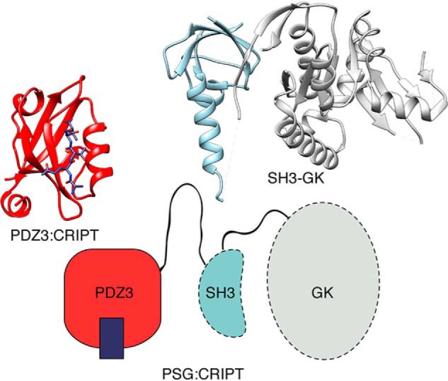
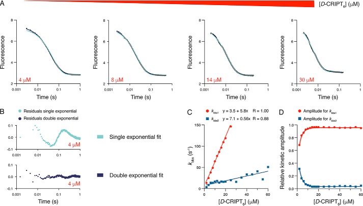
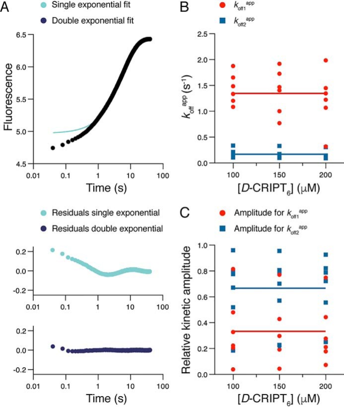
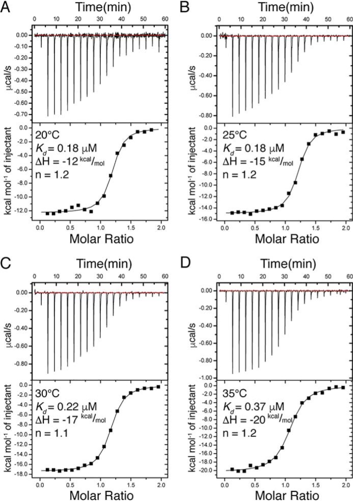
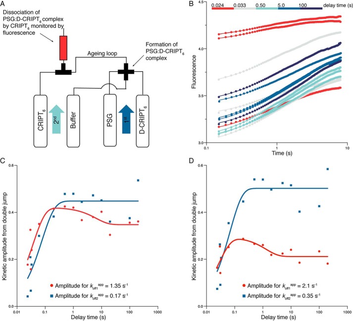
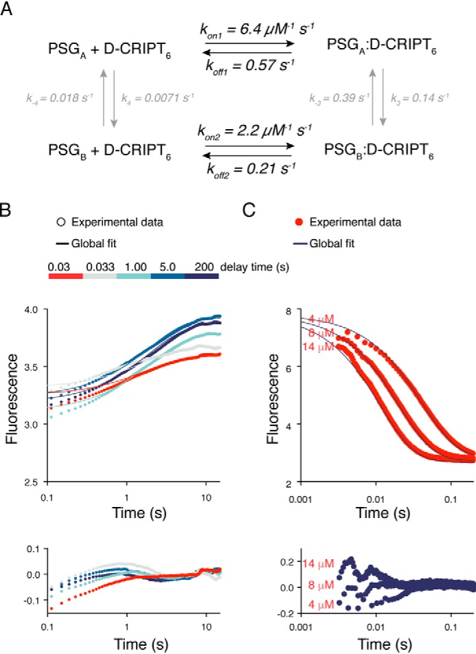
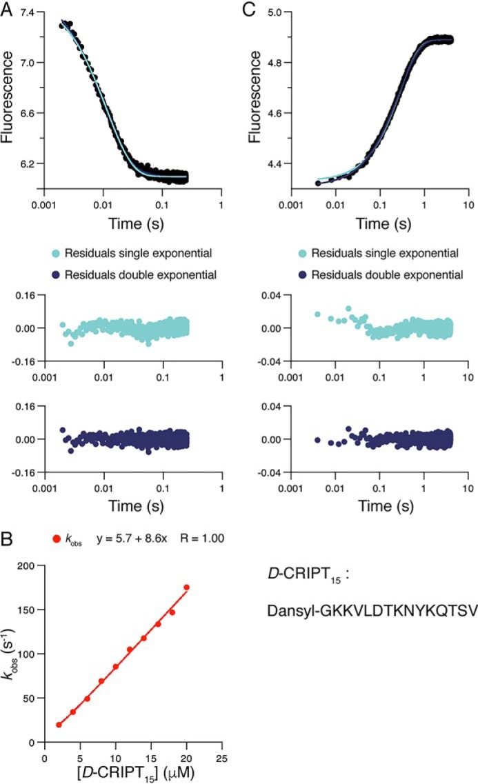
Cell scaffolding and signaling are governed by protein-protein interactions. Although a particular interaction is often defined by two specific domains binding to each other, this interaction often occurs in the context of other domains in multidomain proteins. How such adjacent domains form supertertiary structures and modulate protein-protein interactions has only recently been addressed and is incompletely understood. The postsynaptic density protein PSD-95 contains a three-domain supramodule, denoted PSG, which consists of PDZ, Src homology 3 (SH3), and guanylate kinase-like domains. The PDZ domain binds to the C terminus of its proposed natural ligand, CXXC repeat-containing interactor of PDZ3 domain (CRIPT), and results from previous experiments using only the isolated PDZ domain are consistent with the simplest scenario for a protein-protein interaction; namely, a two-state mechanism. Here we analyzed the binding kinetics of the PSG supramodule with CRIPT. We show that PSG binds CRIPT via a more complex mechanism involving two conformational states interconverting on the second timescale. Both conformational states bound a CRIPT peptide with similar affinities but with different rates, and the distribution of the two conformational states was slightly shifted upon CRIPT binding. Our results are consistent with recent structural findings of conformational changes in PSD-95 and demonstrate how conformational transitions in supertertiary structures can shape the ligand-binding energy landscape and modulate protein-protein interactions.

Keywords: CXXC repeat–containing interactor of PDZ3 domain (CRIPT); PDZ domain; PSD-95; conformational change; kinetics; postsynaptic density protein; protein conformation; protein–protein interaction; supertertiary structure; supramodule.


Conflict of interest statement

The authors declare that they have no conflicts of interest with the contents of this article

Figures

Figure 1.
Figure 1.
Structure of the PSG supramodule from PSD-95. Schematic supertertiary structure of the PSG supramodule from PSD-95 together with crystal structures of PDZ3 with a bound CRIPT peptide (PDB code 1BE9) and of the SH3-GK tandem (PDB code 1JXO).
Figure 2.
Figure 2.
Binding kinetics of the PSG supramodule and CRIPT. A, example of kinetic transients for the association of PSG with D-CRIPT6. PSG (1 μm final concentration) was mixed rapidly in the stopped flow with D-CRIPT6 at different concentrations. B, the residuals are from fits to single- and double-exponential functions, respectively, at 4 μm final concentration of D-CRIPT6. C, the experiment was repeated over a range of D-CRIPT6 concentrations, and the kobs values obtained from the fit to a double-exponential function were plotted versus [D-CRIPT6]. kobs values of more than 150 s−1 were omitted because they very slightly but progressively deviated from a straight line because of instrumental limitations in the mixing. D, kinetic amplitudes associated with the respective kobs value.
Figure 3.
Figure 3.
Dissociation kinetics of the PSG supramodule and D-CRIPT6. A, example of kinetic trace from an experiment where a complex of PSG and D-CRIPT6 (2 and 10 μm) was rapidly mixed with an excess of unlabeled CRIPT (150 μm). The resulting trace was fitted to a single or double exponential, and the residuals from the respective fit are presented below the trace. The double-exponential fit yielded the following parameters: koff1app = 1.35 s−1 (amplitude = 0.32) and koff2app = 0.16 s−1 (amplitude = 1.4). B and C, this experiment was repeated six times, each time at three different concentrations of unlabeled CRIPT (100, 150, and 200 μm). The double-exponential fit yielded koffapp values (B) and their associated amplitudes (C, normalized), which were plotted versus CRIPT concentration. The large scatter in the points, in particular for the amplitudes, reflects the covariation of parameters in the curve fitting. The average of all 18 koffapp values for each kinetic phase yielded the following parameters: koff1app = 1.35 ± 0.42 s−1 and koff2app = 0.17 ± 0.08 s−1 (indicated by horizontal lines in B). The average of all 18 amplitudes yielded the following values: Amp1 = 0.33 ± 0.24 and Amp2 = 0.67 ± 0.24 (indicated by horizontal lines in C).
Figure 4.
Figure 4.
Isothermal titration calorimetry experiments with PSG and D-CRIPT6. A–D, ITC experiments were performed at four different temperatures (20 °C–35 °C). The parameters shown were obtained by fitting a 1:1 binding model using the software provided with the instrument. The Kd values from these experiments agreed well with those calculated from the kinetic experiments at 10 °C (koff1app/kon1 or koff2app/kon2).
Figure 5.
Figure 5.
A kinetic scheme consistent with the binding kinetics of PSG and D-CRIPT6. A scenario with two conformations, PSGA and PSGB, which are in equilibrium and can both bind CRIPT, is consistent with the kinetic and equilibrium data. Access to the binding groove is more restricted in the PSGB conformation.
Figure 6.
Figure 6.
Interrupted binding experiments. A, schematic of the double-jump setup. In the first jump, 4 μm PSG was mixed with 8 μm D-CRIPT6 (i.e. the final concentrations after mixing were 2 μm and 4 μm, respectively) and incubated for a certain delay time in an aging loop. Following the set delay time, any formed complex was dissociated by a large excess of unlabeled CRIPT in a second jump (final concentrations were 1 μm PSG, 2 μm D-CRIPT6, and 50 μm unlabeled CRIPT). B, the dissociation kinetics were monitored in the flow cell. The kinetic traces were fitted simultaneously to a double-exponential function with either locked kobs values (the two average koffapp values from single-jump experiments, 1.35 s−1 and 0.17 s−1, respectively) or shared and free-fitted kobs values (koff1app = 2.1 s−1 and koff2app = 0.35 s−1). The best fit curves in the figure correspond to the latter fit. C and D, the kinetic amplitudes from the respective fits in B were plotted against delay time. This reflects the build up of PSGA:D-CRIPT6 and PSGB:D-CRIPT6, respectively, with time. These amplitude data, in turn, were fitted to a double-exponential function to obtain two “observed double-jump rate constants” for the build up of PSGA and PSGB, respectively. From the fit, kobsDJ values of around 20–40 s−1 and 0.1–0.8 s−1, respectively, were obtained. Although the parameters are underdetermined, it is clear that there is an initial increase in the respective population, followed by a decrease in PSGA:D-CRIPT6. The expected concomitant increase in PSGB:D-CRIPT6 is lost in the experimental noise.
Figure 7.
Figure 7.
Global fitting to a minimal scheme describing PSG:CRIPT binding. Data from binding and dissociation kinetic experiments were fitted globally with KinTek Explorer. A, the scheme shows the fitted square model with the best fit parameters and their standard errors. The amplitude factors for PSGA:D-CRIPT6 and PSGB:D-CRIPT6 were fitted to 0.9 ± 0.1 and 0.13 ± 0.03, respectively. It should be noted that the reported standard errors for all parameters are likely underestimated. We used the global fit to qualitatively test our suggested minimal model. Thus, less emphasis should be put on the estimates of the microscopic rate constants, which are, in some cases, poorly constrained. kon2 is likely overestimated because we could only use lower D-CRIPT6 concentrations in the global fit to capture the fast phase (Fig. 2). B, fit of interrupted binding stopped flow data to the minimal model in A. Five kinetic traces are shown for clarity. The residuals of the fits are shown below the curve. C, fit of the single-jump stopped-flow data to the model, with residuals shown below the curves. The fits to kinetic traces of three different concentrations of D-CRIPT6 are shown for clarity.
Figure 8.
Figure 8.
Binding kinetics of the PSG supramodule and D-CRIPT15. A, PSG (1 μm final concentration) was mixed rapidly in the stopped flow with D-CRIPT15 (10 μm final concentration). A single-exponential function well describes the experimental transient, as shown by comparison of residuals from fit to single- and double-exponential functions. B, the experiment was repeated over a range of D-CRIPT15 concentrations, and kobs obtained from the fit to a single-exponential function was plotted versus [D-CRIPT15] to obtain konapp = (8.4 ± 0.2) × 106 m−1 s−1 (fitting error). C, dissociation of the complex. PSG:D-CRIPT15 (2 and 10 μm) was rapidly mixed with an excess of unlabeled CRIPT (150 μm). The resulting trace was fitted to both single- and double-exponential functions. The residuals from the fits show that the dissociation kinetics follow single exponential kinetics. The average of three experiments using 100, 150, and 200 μm CRIPT, respectively, for displacement yielded koffapp = 3.5 ± 0.011 s−1 (± S.D.).

Similar articles

Cited by

References

    1. Sheng M., and Kim E. (2011) The postsynaptic organization of synapses. Cold Spring Harb. Perspect. Biol. 3, pii: a005678 10.1101/cshperspect.a005678 - DOI - PMC - PubMed
    1. Feng W., and Zhang M. (2009) Organization and dynamics of PDZ-domain-related supramodules in the postsynaptic density. Nat. Rev. Neurosci. 10, 87–99 10.1038/nrn2540 - DOI - PubMed
    1. Ivarsson Y. (2012) Plasticity of PDZ domains in ligand recognition and signaling. FEBS Lett. 586, 2638–2647 10.1016/j.febslet.2012.04.015 - DOI - PMC - PubMed
    1. Tompa P. (2012) On the supertertiary structure of proteins. Nat. Chem. Biol. 8, 597–600 10.1038/nchembio.1009 - DOI - PubMed
    1. Long J.-F., Tochio H., Wang P., Fan J.-S., Sala C., Niethammer M., Sheng M., and Zhang M. (2003) Supramodular structure and synergistic target binding of the N-terminal tandem PDZ domains of PSD-95. J. Mol. Biol. 327, 203–214 10.1016/S0022-2836(03)00113-X - DOI - PubMed

Publication types

MeSH terms

LinkOut - more resources