. 2019 Dec 13;366(6471):eaaw2106.
doi: 10.1126/science.aaw2106.

p27 allosterically activates cyclin-dependent kinase 4 and antagonizes palbociclib inhibition

Affiliations

p27 allosterically activates cyclin-dependent kinase 4 and antagonizes palbociclib inhibition

Keelan Z Guiley et al. Science. .

Abstract

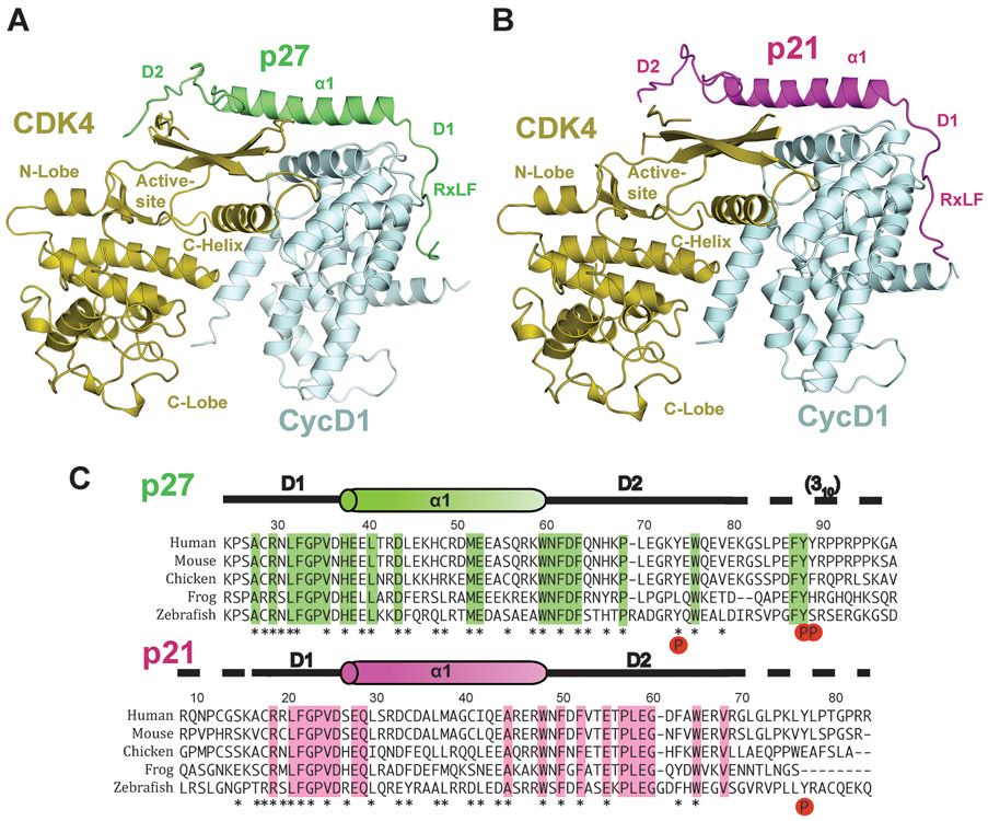
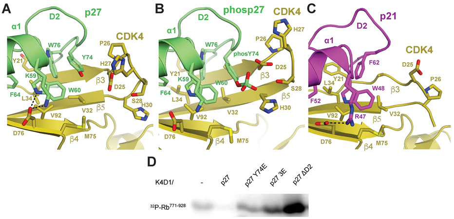
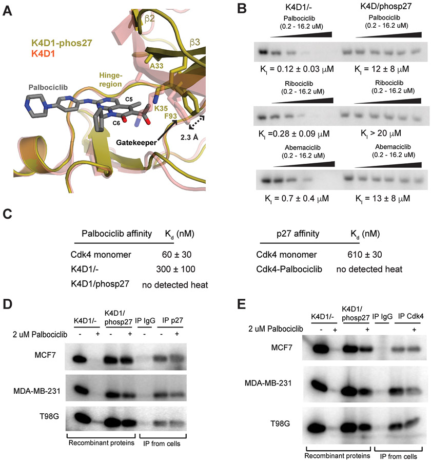
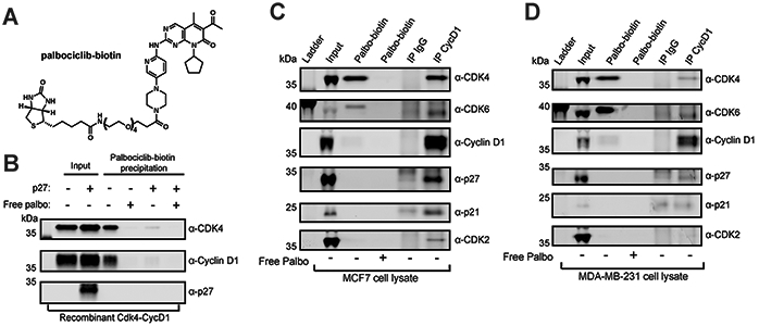
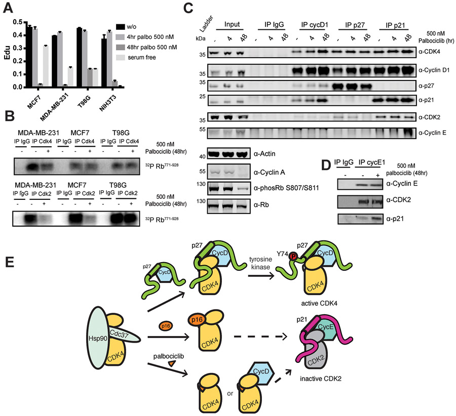
The p27 protein is a canonical negative regulator of cell proliferation and acts primarily by inhibiting cyclin-dependent kinases (CDKs). Under some circumstances, p27 is associated with active CDK4, but no mechanism for activation has been described. We found that p27, when phosphorylated by tyrosine kinases, allosterically activated CDK4 in complex with cyclin D1 (CDK4-CycD1). Structural and biochemical data revealed that binding of phosphorylated p27 (phosp27) to CDK4 altered the kinase adenosine triphosphate site to promote phosphorylation of the retinoblastoma tumor suppressor protein (Rb) and other substrates. Surprisingly, purified and endogenous phosp27-CDK4-CycD1 complexes were insensitive to the CDK4-targeting drug palbociclib. Palbociclib instead primarily targeted monomeric CDK4 and CDK6 (CDK4/6) in breast tumor cells. Our data characterize phosp27-CDK4-CycD1 as an active Rb kinase that is refractory to clinically relevant CDK4/6 inhibitors.


Figures

Fig. 1:
Fig. 1:. Structures of the p27-CDK4-CycD1 and p21-CDK4-CycD1 complexes.
(A) Overall structure of p27-CDK4-CycD1. p27 (green) binds CycD1 (cyan) with its D1 domain and CDK4 (gold) with its D2 domain. (B) Structure of p21-CDK4-CycD1. p21 (magenta) adopts a similar fold to p27, bridging CDK4 (gold) and CycD1 (cyan). (C) Sequence alignment of p27 and p21. Asterisks represent residues directly interacting with CDK4 or CycD1. The known tyrosine phosphorylation sites are noted. Secondary structure observed in the crystal is indicated above the sequences. Dashed lines indicate sequences in the crystallized protein that are not visible in the electron density, including the C-terminal sequence in p27 that forms a 310 helix when bound to CDK2 (in parentheses).
Fig. 2:
Fig. 2:. p27 and p21 inhibit substrate binding and catalytic activity.
(A) Association between the p27 RxLF motif (green) and the MVRIL cleft in CycD1 (cyan) competes for substrate docking. (B) The p21 RxLF (magenta) bound to CycD1 (cyan). (C) Binding of the D2 region in p27 (green) displaces the β1 strand in the CDK4 N-lobe (gold), disrupting the ATP-binding site. The structure shown in grey for comparison, including the ATP, is from CDK2 in the active CDK2-CycA dimer. The CDK4 β1 strand and following Gly-loop are not visible in the p27-CDK4-CycD1 trimer structure, indicating they are disordered. The C-helix remains in an inactive conformation in the CDK4-p27 structure. (D) Comparison of binding of p27 to CDK2 (p27 in grey) and to CDK4 (p27 in green, CDK4 in gold). The 310 helix in p27 that binds the CDK2 ATP-site is not visible and likely remains disordered upon CDK4 binding. Tyrosine phosphorylation sites are shown.
Fig. 3:
Fig. 3:. p27 induces structural changes that promote ATP coordination and processing.
(A-D) Structural alignment of CDK4-CycD1 with p27 (gold-cyan) and without p27 (red, PDB code: 2W96) reveals movement of both the CDK4 N-lobe and CycD1 domains relative to the CDK4 C-lobe. (E) Phosphorylation of purified Rb771-928 with 32P-ATP and the indicated dimer (K4D1/−) or trimer complex. (F-G) Steady state kinase assays measuring effects of ATP concentration on initial reaction rate, measured by incorporation of 32P-ATP. Reactions include CDK4 dimer (K4D1/−) or active trimer (K4D1/phosp27) and the indicated substrate.
Fig. 4:
Fig. 4:. Y74 phosphorylation disrupts the p27 D2-CDK4 interface.
Comparison of CDK4-CycD1 structures with (A) unphosphorylated p27, (B) phosphorylated p27, and (C) p21. (D) 32P-ATP phosphorylation of Rb771-928 using K4D1 dimer or trimer enzymes assembled with the indicted p27 kinase inhibitory domain construct (residues 25-93). 3E contains three glutamate phosphomimetics at Y74, Y88 and Y89. ΔD2 contains p27 residues 25-60, and therefore lacks the D2 CDK4-binding domain
Fig. 5:
Fig. 5:. Palbociclib does not bind and poorly inhibits purified and endogenous CDK4 trimer complexes.
(A) K4D1 dimer (PDB ID: 2W96) and the phosp27-K4D1 trimer structures were aligned with palbociclib-bound CDK6 (PDB ID: 2EUF, not shown) to model the position and interactions of the drug when bound to CDK4. (B) 32P-ATP phosphorylation of Rb771-928 using CDK4-CycD1 dimer (K4D1/−) and phosp27-CDK4-CycD1 trimer (K4D1/phosp27) enzymes in the absence (left most lane in each titration) and presence of increasing inhibitor concentrations (see Fig. S8A for quantification). Each drug is dosed from 0.2 μM to 16.2 μM in 3-fold increments. (C) ITC affinities for palbociclib (left) or p27 (right) titrated into the indicated enzyme. (D) The indicated cell lysates were immunoprecipitated with control or with p27 antibody, and the activity of the immunoprecipitate was used to phosphorylate Rb771-928 with 32P-ATP in the absence or presence of palbociclib. Reactions with the indicated recombinant dimer (K4D1/−) or trimer (K4D1/phosp27) enzymes are shown for comparison in the first four lanes. (E) As in panel D, except lysates were precipitated with antiserum raised against a CDK4 C-terminal peptide.
Fig. 6:
Fig. 6:. The ATP-site occupancy probe XO44 labels monomer CDK4 in MCF7 cells.
(A) Labeling of endogenous CDK4 and CDK2 by the promiscuous covalent ATP-site probe XO44. MCF7 cells were treated with DMSO vehicle or XO44 at the indicated concentrations for 30 min. Lysates were subjected to click reaction with TAMRA–azide to visualize XO44 labeling of proteins by gel mobility shift. (B) Palbociclib competes with XO44 for CDK4 binding but not CDK2 binding. Experiment performed as in panel A, but cells were pretreated for 60 mins with DMSO, a non-clickable analog of XO44 (XO-nc), or with increasing concentrations of palbociclib. (C) XO44 efficiently labels purified recombinant CDK4-CycD1 dimer but not trimer complexes with p21 or p27 as determined by electrospray ionization mass spectrometry. Protein complexes were treated with DMSO or XO44. Average percent labeling was determined for three replicates with standard deviation shown as error bars. (D) Asynchronous MCF7 cells were treated with DMSO, XO44 (2 μM, 30 min) or palbociclib (500 nM, 4 hr), and lysates were fractionated using Superdex200 size-exclusion chromatography. XO44 labeling was monitored by gel mobility shift after click reaction with TAMRA–N3 (“XO44 click”). Normalized quantification of band signals from the western blots using the indicated antibody are shown. A benchmark chromatography experiment using the recombinant protein complex is displayed at the bottom of the panel. See Fig. S10 for data using MDA-MB-231 cells.
Fig. 7:
Fig. 7:. Palbociclib directly targets CDK4/6 monomer in cell lysate.
(A) Chemical structure of palbociclib-biotin synthesized here. (B) Palbociclib-biotin precipitated purified recombinant CDK4-CycD1 dimer, but not p27-CDK4-CycD1 trimer. (C and D) MCF7 or MDA-MB-231 cell lysates were used in a precipitation reaction with palbociclib-biotin. A CycD1 antibody was also used to precipitate CDK4/6 from cell lysates to compare the total pool of CycD1 complexes to palbociclib-bound complexes.
Fig. 8:
Fig. 8:. Palbociclib indirectly leads to down-regulation of CDK2 through p21.
(A) Cell-cycle profiling of the cell lines used in this study upon treatment with 500 nM palbociclib for 4 or 48 hr or without drug treatment. Cells were assayed for 5-ethynyl-2-deoxyuridine (EdU) incorporation as a marker of S phase at the different time points following drug treatment. The fraction of cells showing EdU staining is reported. EdU incorporation in cells that were serum starved (serum free) for 48 hours is also shown for comparison. (B) Cells were treated with palbociclib as indicated for 48 hr, and lysates were immunoprecipitated with CDK4 antiserum or a CDK2 antibody. Activity of the immunoprecipitated complexes was assayed as in Fig. 5, D and E. (C) CycD1-, p27-, and p21-associated complexes were immunoprecipitated from MCF7 cells treated with palbociclib for the indicated time and proteins were detected by Western blot. (D) CycE1 was immunoprecipitated from MCF7 cells treated with palbociclib for 48 hrs. (E) Model for trimer assembly and CDK4 inhibition by palbociclib. Like p16 family CDK4 inhibitors, palbociclib binds monomer CDK4 and indirectly leads to inactive CDK2 complexes. Although not observed in the breast cancer cells studied here, there may be some contexts in which palbociclib also targets CDK4 dimers.

Comment in

References

    1. Hunter T, Pines J, Cyclins and cancer. II: Cyclin D and CDK inhibitors come of age. Cell 79, 573–582 (1994). - PubMed
    1. Sherr CJ, Cancer cell cycles. Science 274, 1672–1677 (1996). - PubMed
    1. Malumbres M, Cyclin-dependent kinases. Genome Biol 15, 122 (2014). - PMC - PubMed
    1. Otto T, Sicinski P, Cell cycle proteins as promising targets in cancer therapy. Nat Rev Cancer 17, 93–115 (2017). - PMC - PubMed
    1. Sherr CJ, Beach D, Shapiro GI, Targeting CDK4 and CDK6: From Discovery to Therapy. Cancer Discov 6, 353–367 (2016). - PMC - PubMed

Publication types

MeSH terms