. 2019 Dec 12;9(1):18916.
doi: 10.1038/s41598-019-55388-0.

Ultrasound-induced Cavitation enhances the efficacy of Chemotherapy in a 3D Model of Pancreatic Ductal Adenocarcinoma with its microenvironment

Affiliations

Ultrasound-induced Cavitation enhances the efficacy of Chemotherapy in a 3D Model of Pancreatic Ductal Adenocarcinoma with its microenvironment

R Leenhardt et al. Sci Rep. .

Abstract

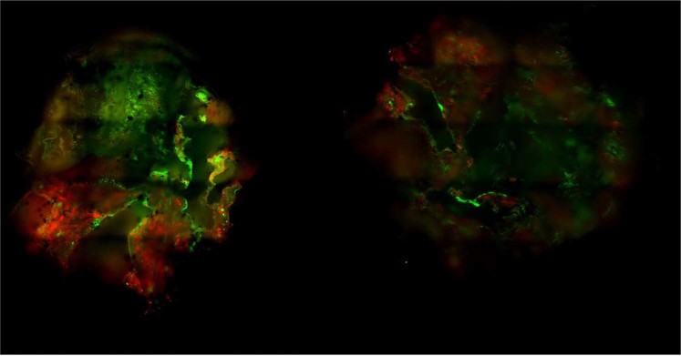
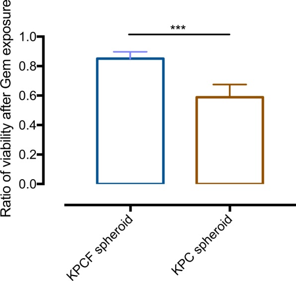
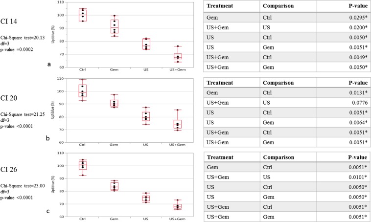
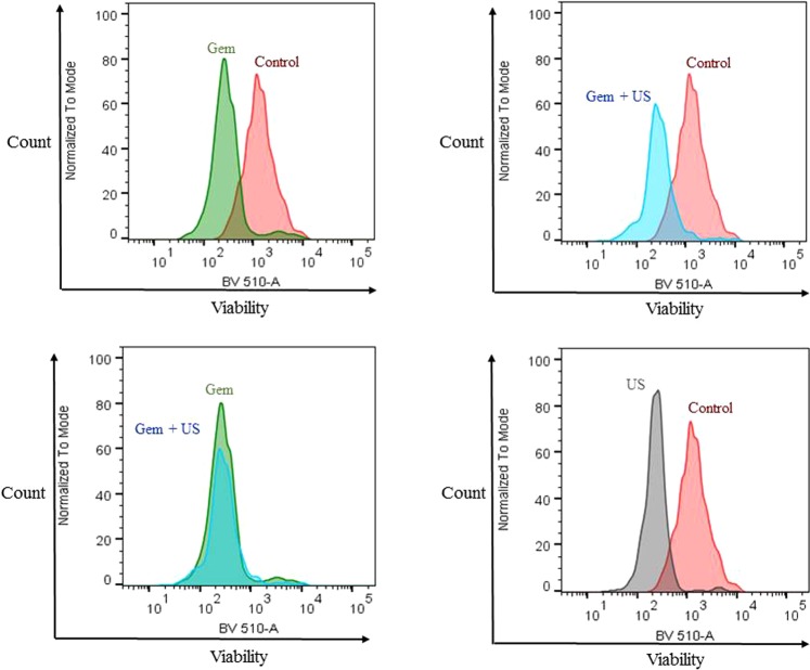
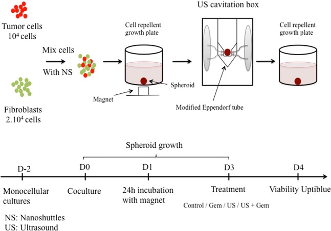
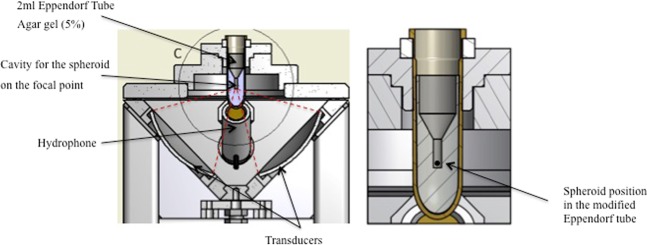
Pancreatic ductal adenocarcinoma (PDAC) is supported by a complex microenvironment whose physical contribution to chemoresistance could be overcome by ultrasound (US) therapy. This study aims to investigate the ability of US-induced inertial cavitation in association with chemotherapy to alter tumor cell viability via microenvironment disruption. For this purpose, we used a 3D-coculture PDAC model partially mimicking the tumor and its microenvironment. Coculture spheroids combining DT66066 cells isolated from KPC-transgenic mice and murine embryonic fibroblasts (iMEF) were obtained by using a magnetic nanoshuttle method. Spheroids were exposed to US with incremental inertial cavitation indexes. Conditions studied included control, gemcitabine, US-cavitation and US-cavitation + gemcitabine. Spheroid viability was assessed by the reduction of resazurin and flow cytometry. The 3D-coculture spheroid model incorporated activated fibroblasts and produced type 1-collagen, thus providing a partial miniature representation of tumors with their microenvironment. Main findings were: (a) Gemcitabine (5 μM) was significantly less cytotoxic in the presence of KPC/iMEFs spheroids compared with KPC (fibroblast-free) spheroids; (b) US-induced inertial cavitation combined with Gemcitabine significantly decreased spheroid viability compared to Gemcitabine alone; (c) both cavitation and chemotherapy affected KPC cell viability but not that of fibroblasts, confirming the protective role of the latter vis-à-vis tumor cells. Gemcitabine toxicity is enhanced when cocultured spheroids of KPC and iMEF are exposed to US-cavitation. Although the model used is only a partial representation of PDAC, this experience supports the hypothesis that US-inertial cavitation can enhance drug penetration and cytotoxicity by disrupting PDAC microenvironment.


Conflict of interest statement

The authors declare no competing interests.

Figures

Figure 1
Figure 1
Immuno-fluorescence imaging of KPCF spheroid at day 3 of growth. KPC and iMEFs respectively expressed mCherry (red) and Green Fluorescent Protein (GFP) for clear immuno-fluorescent demarcation of both components.
Figure 2
Figure 2
Histological section (5 microns) of a KPCF spheroid after 10 days of incubation. Staining with HE (Hematoxylin Eosin) and Masson’s Trichrome. Collagen fibers appear in blue. Blackish deposits correspond to residual magnetic nanoparticles.
Figure 3
Figure 3
Ratio of viability between KPCF and KPC spheroids treated with Gem compared to control. Viability was measured 24 h after low-dose (5 μM) Gemcitabine incubation: Comparison of KPCF (KPC + fibroblasts) vs KPC (monotypic) spheroid models (n = 6 spheroids for each condition).
Figure 4
Figure 4
KPCF viability after various treatment conditions: Gem/US/US + Gem. (a) KPCF viability after US CI 14 treatment combined or not to Gem exposure. N = 6 spheroids for each condition. (b) KPCF viability after US CI 20 treatment combined or not to Gem exposure. N = 6 spheroids for each condition. (c) KPCF viability after US CI 26 treatment combined or not to Gem exposure. N = 6 spheroids for each condition.
Figure 5
Figure 5
KPCF spheroid viability after US CI 20 treatment and Gem exposure. Cytometer analysis. Analysis based on 4 repeat experiments with 6 spheroids pooled for each condition.
Figure 6
Figure 6
Example of the cytometer analysis assessing the viability measured by BV 510-A of KPC cells from dissociated KPCF spheroids after various treatments.
Figure 7
Figure 7
Coculture protocol/Formation of tumor spheroids/Treatment protocol design.
Figure 8
Figure 8
Spheroid position in the modified Eppendorf® tube.

Similar articles

Cited by

References

    1. Lucas AL, et al. Global Trends in Pancreatic Cancer Mortality From 1980 Through 2013 and Predictions for 2017. Clin. Gastroenterol. Hepatol. Off. Clin. Pract. J. Am. Gastroenterol. Assoc. 2016;14:1452–1462.e4. - PMC - PubMed
    1. Sultana A, et al. Systematic review, including meta-analyses, on the management of locally advanced pancreatic cancer using radiation/combined modality therapy. Br. J. Cancer. 2007;96:1183–1190. doi: 10.1038/sj.bjc.6603719. - DOI - PMC - PubMed
    1. Ryan DP, Hong TS, Bardeesy N. Pancreatic adenocarcinoma. N. Engl. J. Med. 2014;371:1039–1049. doi: 10.1056/NEJMra1404198. - DOI - PubMed
    1. Maitra A, Hruban RH. Pancreatic cancer. Annu. Rev. Pathol. 2008;3:157–188. doi: 10.1146/annurev.pathmechdis.3.121806.154305. - DOI - PMC - PubMed
    1. Olive KP, et al. Inhibition of Hedgehog signaling enhances delivery of chemotherapy in a mouse model of pancreatic cancer. Science. 2009;324:1457–1461. doi: 10.1126/science.1171362. - DOI - PMC - PubMed

Publication types

MeSH terms