. 2019 Dec 12;9(1):18942.
doi: 10.1038/s41598-019-55405-2.

Tensin1 expression and function in chronic obstructive pulmonary disease

Affiliations

Tensin1 expression and function in chronic obstructive pulmonary disease

Panayiota Stylianou et al. Sci Rep. .

Abstract

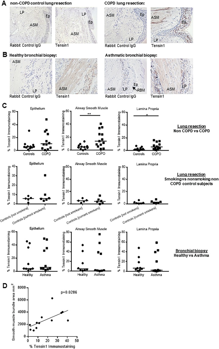
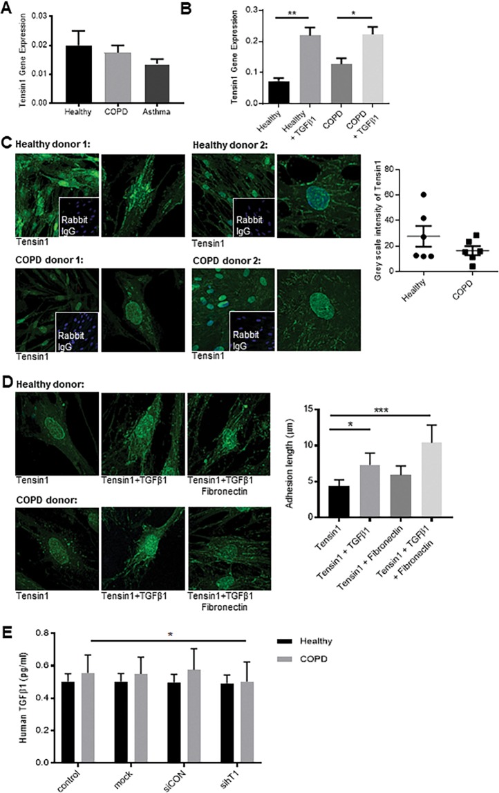
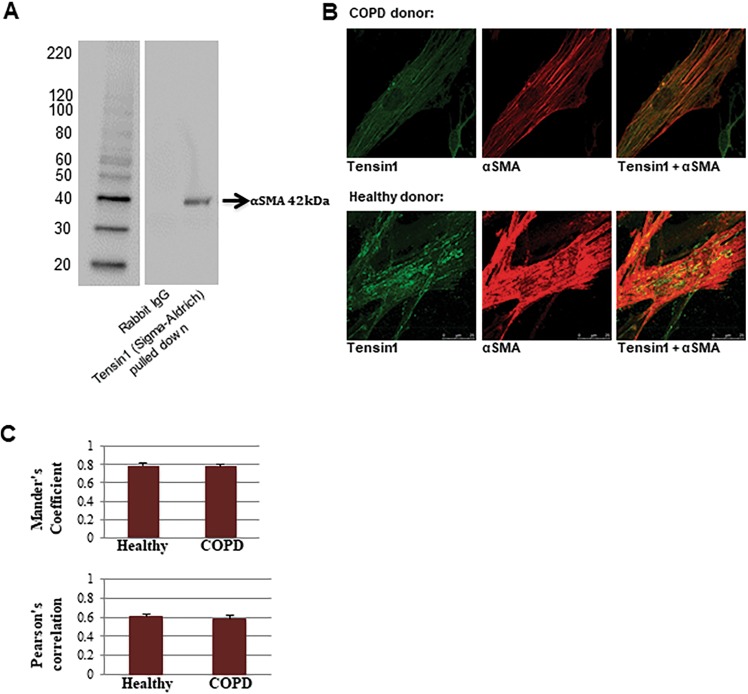
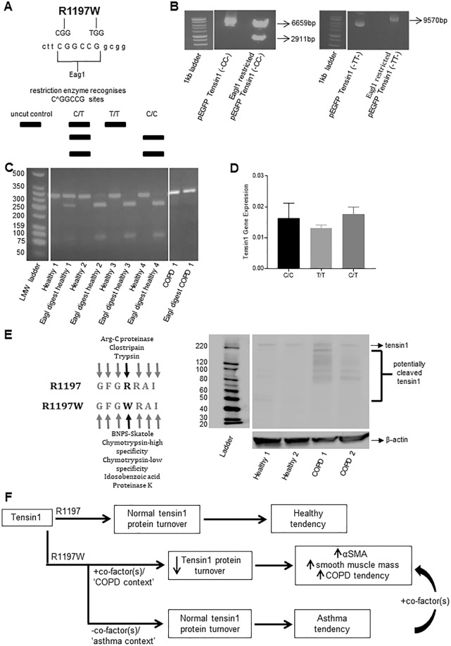
Chronic obstructive pulmonary disease (COPD) constitutes a major cause of morbidity and mortality. Genome wide association studies have shown significant associations between airflow obstruction or COPD with a non-synonymous SNP in the TNS1 gene, which encodes tensin1. However, the expression, cellular distribution and function of tensin1 in human airway tissue and cells are unknown. We therefore examined these characteristics in tissue and cells from controls and people with COPD or asthma. Airway tissue was immunostained for tensin1. Tensin1 expression in cultured human airway smooth muscle cells (HASMCs) was evaluated using qRT-PCR, western blotting and immunofluorescent staining. siRNAs were used to downregulate tensin1 expression. Tensin1 expression was increased in the airway smooth muscle and lamina propria in COPD tissue, but not asthma, when compared to controls. Tensin1 was expressed in HASMCs and upregulated by TGFβ1. TGFβ1 and fibronectin increased the localisation of tensin1 to fibrillar adhesions. Tensin1 and α-smooth muscle actin (αSMA) were strongly co-localised, and tensin1 depletion in HASMCs attenuated both αSMA expression and contraction of collagen gels. In summary, tensin1 expression is increased in COPD airways, and may promote airway obstruction by enhancing the expression of contractile proteins and their localisation to stress fibres in HASMCs.


Conflict of interest statement

The authors declare no competing interests.

Figures

Figure 1
Figure 1
Tensin1 immunostaining in non-disease controls and COPD lung resection and healthy and asthmatic bronchial biopsies. (A) Examples of tensin1 and isotype control immunostaining in COPD (n = 13) and non-COPD lung resections (n = 11) (EP: epithelium; ASM: airway smooth muscle; LP: lamina propria). (B) Examples of tensin1 and isotype control immunostaining in bronchial biopsies from patients with asthma (n = 10) and healthy controls (n = 9) (EP: epithelium; ASM: airway smooth muscle; LP: lamina propria, RBM: reticular basement membrane). (C) The extent of tensin1 immunostaining in airway epithelium, airway smooth muscle and lamina propria analysed using threshold measurements. Increased tensin1 immunostaining was observed in the airway smooth muscle (**p = 0.007) and lamina propria (*p = 0.012) in COPD subjects when compared to non-COPD controls (Mann-Whitney unpaired non parametric test). Tensin1 immunostaining in lung resections was not affected by the smoking status of non-COPD control individuals. (D) Tensin1 immunostaining in COPD subjects was positively correlated with the smooth muscle bundle area, calculated by Pearson’s correlation (R = 0.6286, p = 0.0286).
Figure 2
Figure 2
Tensin1 mRNA and protein expression in HASMCs in health and disease and the effect of TGFβ1 and fibronectin. (A) Quantitative Reverse Transcription -PCR (qRT-PCR) was used to quantify tensin1 mRNA expression in HASMCs isolated from healthy donors and patients with COPD or asthma using the 2^-(ΔCt) method (n = 7 of each, Mean ± SEM). (B) Tensin1 mRNA expression in human airway smooth muscle cells (HASMCs) was increased following TGFβ1-dependent stimulation in both COPD (n = 3) (*p = 0.0418) and healthy (n = 3) (**p = 0.0042) donors (Tukey’s multiple comparison test as part of one-way ANOVA, one-way ANOVA test: **p = 0.0019). (C) HASMC tensin1 immunofluorescent staining was measured by grey scale intensity in n = 6 healthy and n = 6 COPD subjects. No difference was observed in the grey scale intensity of tensin1 immunostaining in the two phenotypes (Mean ± SEM). (D) Cells were stimulated with TGFβ1 and/or fibronectin. Stimulation with TGFβ1 and fibronectin did not significantly increase the grey scale intensity of tensin1. However, the length of fibrillar adhesions was significantly increased in HASMCs stimulated with TGFβ1 alone (*p = 0.0386) and TGFβ1 + fibronectin (***p = 0.0002) (Dunnett’s multiple comparison test as part of one-way ANOVA, one-way ANOVA test: ***p = 0.0004). Data shown is pooled COPD and healthy donors which did not differ (n = 3 of each, Mean ± SEM). (E) Cells were transfected with siRNA directed against tensin1 and supernatants were assessed for TGFβ1 expression using ELISA in both COPD (n = 4) and healthy donors (n = 4) (Mean ± SEM). Tensin1 depleted HASMC supernatants derived from COPD subjects had a small but significant reduction of TGFβ1 secretion when compared to controls (*p = 0.0477) (paired t-test).
Figure 3
Figure 3
Tensin1 co-localises and correlates with αSMA in HASMCs. (A) Co-immunoprecipitation was carried out to investigate the interaction of tensin1 and αSMA. Tensin1 immunoprecipitates were analysed by western blotting analysis using an αSMA antibody. A band of 42 kDa was detected suggesting a physical interaction between tensin1 and αSMA. (B) Confocal immunofluorescent staining demonstrating co-localisation of tensin1 (green) and αSMA (red) in HAMSCs. (C) Mander’s overlap coefficient and Pearson’s correlation were calculated to confirm association of the two proteins on n = 3 healthy and n = 3 COPD subjects (Mean ± SEM).
Figure 4
Figure 4
Reduced αSMA mRNA and protein expression in tensin1-depleted HASMCs. Tensin1 siRNA-transfected and control (untreated cells, transfection reagent alone, non-targeting siRNA control) HASMCs derived from healthy individuals and COPD patients were analysed for αSMA mRNA and protein expression. Cells were also stimulated with TGFβ1 to examine its role in αSMA expression after silencing tensin1. (A) The effects of depleting tensin1 on HASMC αSMA mRNA expression was examined using qRT-PCR on n = 4 COPD donors. αSMA mRNA expression was quantified using the 2−(ΔCt) method (Mean ± SEM). Silencing of tensin1 resulted in significant downregulation of αSMA mRNA in HASMCs when compared to control (p = 0.0011 by repeated measures ANOVA). *p < 0.05, **p < 0.01 by Sidaks multiple comparison test (Mean ± SEM). (B) Cells were transfected with siRNA directed against tensin1 and assessed for αSMA expression using immunofluorescence analysis. Tensin1-depleted HASMCs had a significantly lower intensity of αSMA when compared to controls both in the absence or presence of TGFβ1 (p = 0.0002 by repeated measures ANOVA). **p < 0.05, ** p < 0.01, *** p < 0.001 by Sidaks multiple comparison test. Mean ± SEM. Data in B are pooled COPD (n = 3) and healthy donors (n = 3) which did not differ. (C) The effects of depleting tensin1 on HASMC αSMA protein expression was also confirmed using western blot analysis (p = 0.0006 by repeated measures ANOVA). *p < 0.05, **p < 0.01 by Sidaks multiple comparison test. Mean ± SEM. The left panel shows a representative western blot. Data in the right panel are pooled COPD (n = 4) and healthy donors (n = 4) which did not differ.
Figure 5
Figure 5
Collagen gel contraction by HASMC is dependent on tensin1. (A) Cells were transfected with siRNA directed against tensin1 and incubated within 3D collagen gels. The extent of spontaneous collagen gel contraction was recorded at 4, 18, 24 and 48 hours. Quantification of collagen gel contraction using gel area measurement was performed (n = 8, Mean ± SEM). Data shown are pooled COPD (n = 4) and healthy donors (n = 4) which did not differ. HASMCs transfected with tensin1 siRNA SMARTpool showed a greatly reduced ability to contract, with significant differences compared to controls at 18 (*p = 0.0118), 24 (**p = 0.0027) and 48 hours (****p = 0.0001) (Dunnett’s multiple comparison test as part of two-way ANOVA). (B) Cells were stimulated with bradykinin and collagen gel contraction was assessed on COPD (n = 4) and healthy donors (n = 4) (Mean ± SEM) after 18 hours. Contraction in tensin1 depleted HASMCs was greatly reduced (**p = 0.0033). The extent of collagen gel contraction following bradykinin stimulation was significantly increased in non- (***p = 0.0009), mock (***p = 0.0009) and siCON (***p = 0.0001) transfected cells, when compared to spontaneous contraction. Tensin1-depleted HASMCs did not respond significantly to bradykinin, when compared to controls (Tukey’s multiple comparison test as part of two-way ANOVA).
Figure 6
Figure 6
The distribution of C and T alleles generating amino acid 1997 of tensin1 and schematic illustrating potential role of tensin1 in disease. (A) A diagram illustrating the base substitution at the site of the variant nucleotide base, the Eag1 restriction site formation in the presence of C versus T, and the different tensin1 genotypes evident on digested PCR products. (B) pEGFP-C1 constructs with the tensin1 variants were used as controls to confirm validity of the RFLP technique. (C) An agarose gel image illustrating the different tensin1 phenotypes in HAMSCs. Lane 1 = ladder. Lane 3 = Heterozygous, Lane 5, 7 and 9 = homozygous (-CC-) (encodes R1197 tensin1) and Lane 11 = homozygous (-TT-) (encodes R1197W tensin1). (D) Tensin1 mRNA expression was assessed between the different genotypes that segregated with health and disease. (E) A western blotting analysis illustrating differential cleavage of tensin1 in COPD subjects when compared to healthy controls (left). ‘PeptideCutter’ prediction tool showing differential cleavage at the site of mutation (right). (F) A schematic illustrating our hypothesis that the R1197W mutation and COPD-specific co-factors together modulate tensin1 degradation and thereby affect αSMA expression, and ASM mass.

References

    1. Yang Q, et al. NO and EDHF pathways in pulmonary arteries and veins are impaired in COPD patients. Vascul Pharmacol. 2012;57:113–118. doi: 10.1016/j.vph.2012.05.004. - DOI - PubMed
    1. Shaykhiev R, Crystal RG. Innate immunity and chronic obstructive pulmonary disease: a mini-review. Gerontology. 2013;59:481–489. doi: 10.1159/000354173. - DOI - PMC - PubMed
    1. Zhang J, He J, Xia J, Chen Z, Chen X. Delayed apoptosis by neutrophils from COPD patients is associated with altered Bak, Bcl-xl, and Mcl-1 mRNA expression. Diagn Pathol. 2012;7:65. doi: 10.1186/1746-1596-7-65. - DOI - PMC - PubMed
    1. Decramer M, Janssens W, Miravitlles M. Chronic obstructive pulmonary disease. Lancet. 2012;379:1341–1351. doi: 10.1016/s0140-6736(11)60968-9. - DOI - PMC - PubMed
    1. Pauwels RA, Buist AS, Calverley PM, Jenkins CR, Hurd SS. Global strategy for the diagnosis, management, and prevention of chronic obstructive pulmonary disease. NHLBI/WHO Global Initiative for Chronic Obstructive Lung Disease (GOLD) Workshop summary. Am J Respir Crit Care Med. 2001;163:1256–1276. doi: 10.1164/ajrccm.163.5.2101039. - DOI - PubMed

Publication types