. 2019 Dec 12;9(1):19002.
doi: 10.1038/s41598-019-55567-z.

A novel application of Gini coefficient for the quantitative measurement of bacterial aggregation

Affiliations

A novel application of Gini coefficient for the quantitative measurement of bacterial aggregation

Yu-Ming Cai et al. Sci Rep. .

Abstract

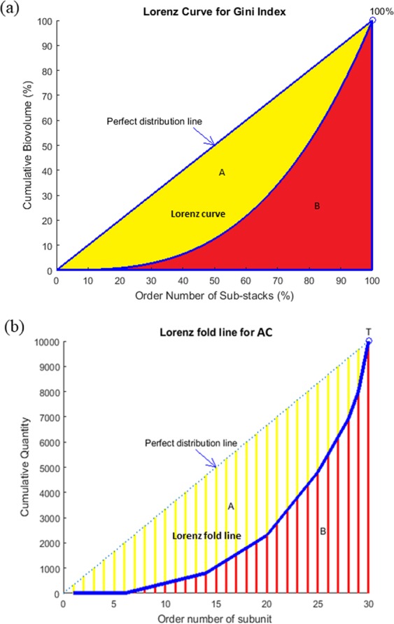
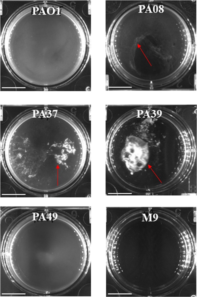
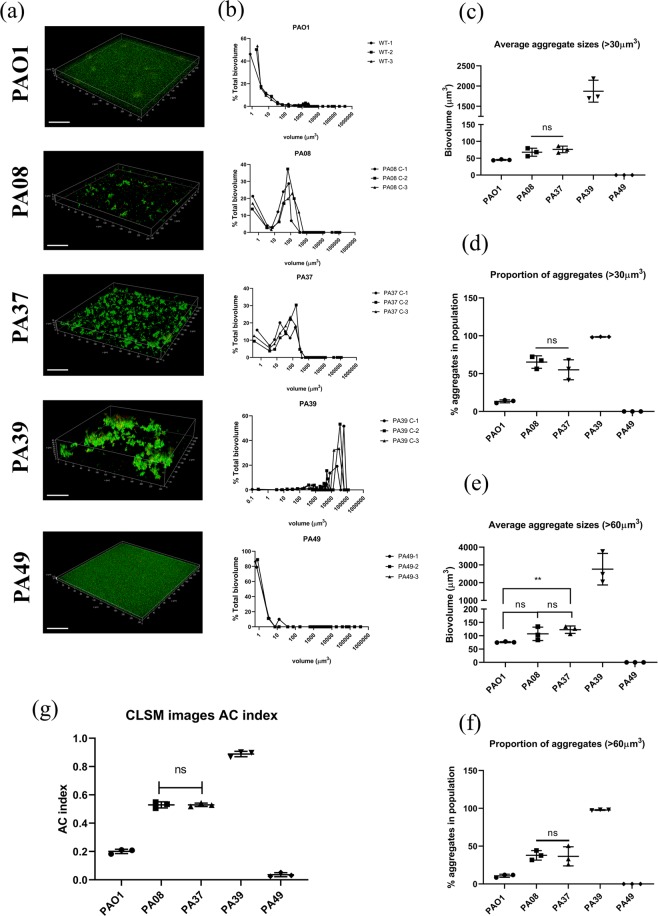
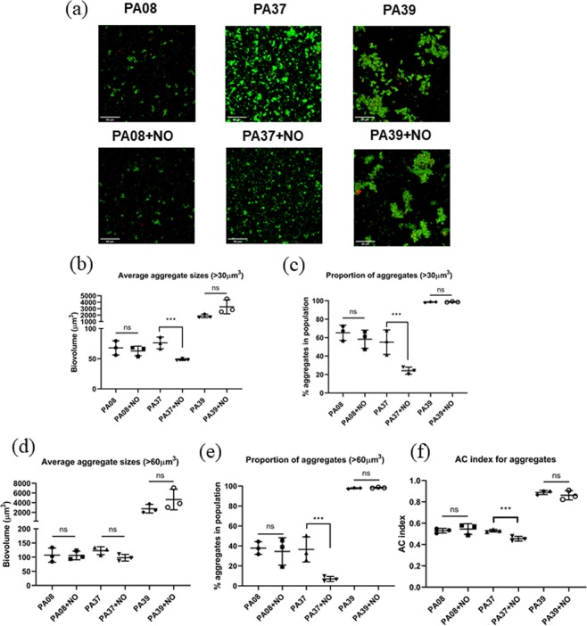
Non-surface attached bacterial aggregates are frequently found in clinical settings associated with chronic infections. Current methods quantifying the extent to which a suspended bacterial population is aggregated mainly rely on: (1) cell size distribution curves that are difficult to be compared numerically among large-scale samples; (2) the average size/proportion of aggregates in a population that do not specify the aggregation patterns. Here we introduce a novel application of Gini coefficient, herein named Aggregation Coefficient (AC), to quantify the aggregation levels of cystic fibrosis Pseudomonas aeruginosa (CF-PA) isolates in vitro using 3D micrographs, Fiji and MATLAB. Different aggregation patterns of five strains were compared statistically using the numerical AC indexes, which correlated well with the size distribution curves plotted by different biovolumes of aggregates. To test the sensitivity of AC, aggregates of the same strains were treated with nitric oxide (NO), a dispersal agent that reduces the biomass of surface attached biofilms. Strains unresponsive to NO were reflected by comparable AC indexes, while those undergoing dispersal showed a significant reduction in AC index, mirroring the changes in average aggregate sizes and proportions. Therefore, AC provides simpler and more descriptive numerical outputs for measuring different aggregation patterns compared to current approaches.


Conflict of interest statement

The authors declare no competing interests.

Figures

Figure 1
Figure 1
Schematic diagrams of different cell distributions in micrographs with the same total number of cells. (a) Cells distribute evenly (equidistribution), AC = 0. (b) Cells distribution is inhomogeneous, AC = 0.533 (c) Cells distribution is very concentrated, AC = 1.
Figure 2
Figure 2
(a) Lorenz curve for a standard Gini coefficient (b) Modified Lorenz fold line for aggregation coefficient.
Figure 3
Figure 3
24 hrs (a) and 72 hrs (b) CF-PA isolates biofilms in M9 medium in microtiter plates. The biofilm formation of each CF-PA strain is compared to PAO1 using two-tailed Student T test. *** denotes P < 0.01, ** denotes 0.01 < P < 0.05. Data acquired from 3 independent experiments and 6 technical replicates.
Figure 4
Figure 4
CF-PA suspended bacterial aggregates in M9 medium cultured in 6 well plates. Red arrows point at macro-aggregates. Scale bar = 1 cm.
Figure 5
Figure 5
(a) The 3D micrographs of cell aggregates imaged by CLSM. Data acquired from 3 independent experiments. Scale bar = 50 µm. (b) Particle biovolumes distribution of different CF-PA planktonic cultures. The sizes of single cells and aggregates are grouped into 26 volume categories. The Sum and Mean biovolumes of particles in each category were calculated. For each category, the proportion of Sum biovolume in the total biovolume of entire micrograph is plotted to the Mean size. The details for each replicate was shown in the same graph to show the variances. (c) Average aggregate biovolume (>30 μm3) in each sample. (d) The proportion of all aggregates (>30 μm3) in the total biovolume of each sample. (e) Average aggregate biovolume (>60 μm3) in each sample. (f) The proportion of all aggregates (>60 μm3) in the total biovolume of each sample. (g) AC indexes of different CF-PA strains in planktonic cultures.
Figure 6
Figure 6
(a) The maximum projection micrographs of cell aggregates. Scale bar = 50 µm. (b) Average aggregate biovolume (>30 μm3) in each sample. (c) The proportion of all aggregates (>30 μm3) in the total biovolume of each sample. (d) Average aggregate biovolume (>60 μm3) in each sample. (e) The proportion of all aggregates (>60 μm3) in the total biovolume of each sample. (f) AC indexes of PA08, PA37 and PA39 suspended aggregates with and without NO treatment. *** denotes P < 0.01, ** denotes P < 0.05. Data acquired from 3 independent experiments.

References

    1. Bjarnsholt T, et al. The in vivo biofilm. Trends in Microbiology. 2013;21:466–74. doi: 10.1016/j.tim.2013.06.002. - DOI - PubMed
    1. Alhede M, et al. Phenotypes of non-attached Pseudomonas aeruginosa aggregates resemble surface attached biofilm. PLoS One. 2011;6:e27943. doi: 10.1371/journal.pone.0027943. - DOI - PMC - PubMed
    1. Kirketerp-Møller K, et al. Distribution, organization, and ecology of bacteria in chronic wounds. J. Clin. Microbiol. 2008;46:2717–2722. doi: 10.1128/JCM.00501-08. - DOI - PMC - PubMed
    1. Quijano G, Arcila JS, Buitrón G. Microalgal-bacterial aggregates: Applications and perspectives for wastewater treatment. Biotechnology Advances. 2017;35:772–781. doi: 10.1016/j.biotechadv.2017.07.003. - DOI - PubMed
    1. Bay L, et al. Bacterial aggregates establish at the edges of acute epidermal wounds. Adv. Wound Care. 2018;7:105–113. doi: 10.1089/wound.2017.0770. - DOI - PMC - PubMed

Publication types