. 2019 Dec 7;25(45):6634-6652.
doi: 10.3748/wjg.v25.i45.6634.

Toxoplasma ROP16I/III ameliorated inflammatory bowel diseases via inducing M2 phenotype of macrophages

Affiliations

Toxoplasma ROP16I/III ameliorated inflammatory bowel diseases via inducing M2 phenotype of macrophages

Yong-Wei Xu et al. World J Gastroenterol. .

Abstract

Background: Inflammatory bowel disease (IBD) is characterized by chronic and non-specific inflammation of the intestinal mucosa and mainly includes ulcerative colitis and Crohn's disease.

Aim: To explore the beneficial effect of ToxoROP16I/III-induced M2 phynotype macrophages in homeostasis of IBDs through downregulation of M1 inflammatory cells.

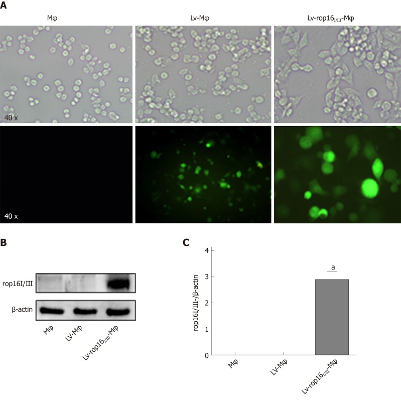
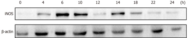
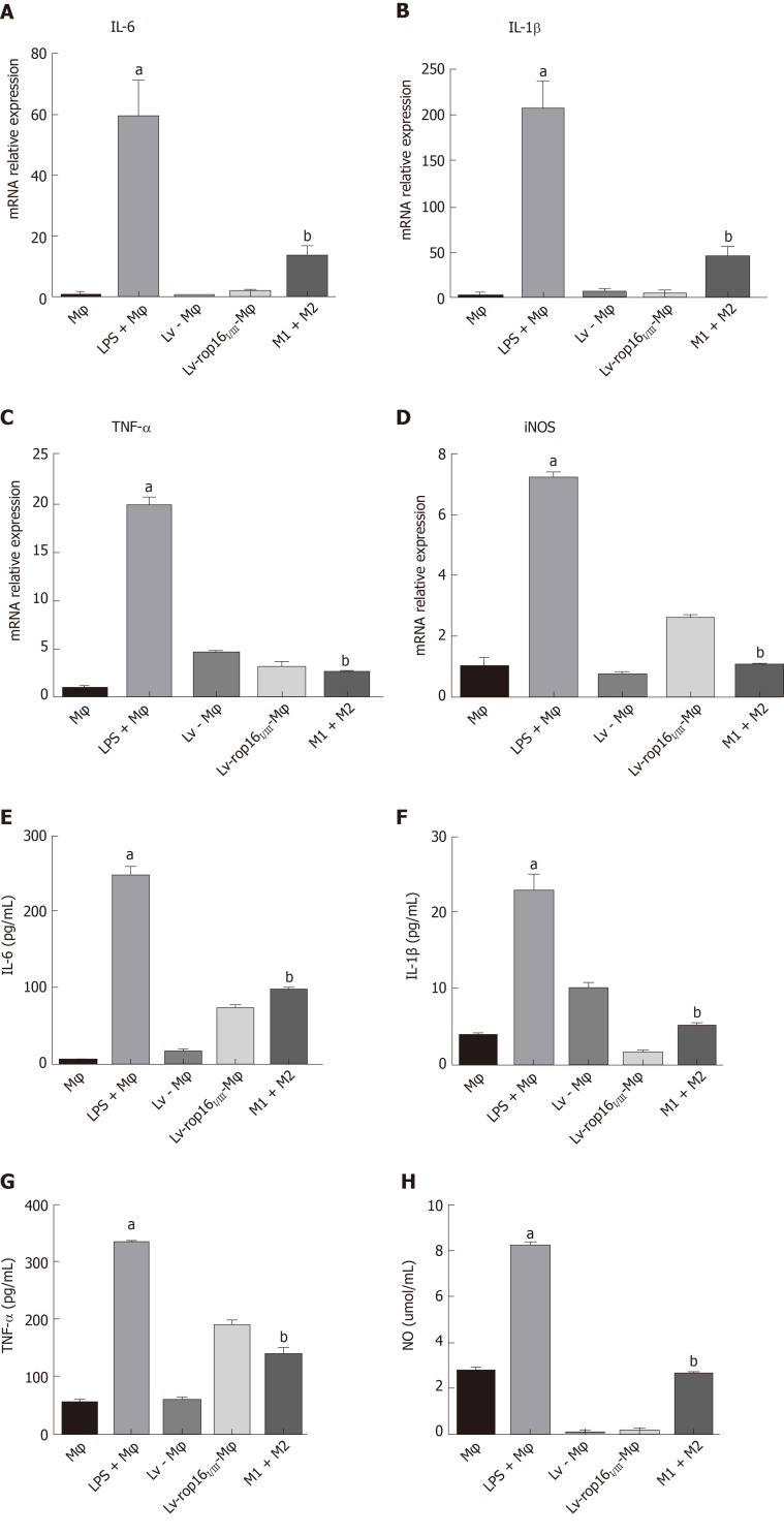
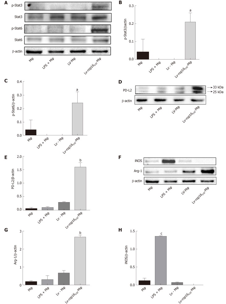
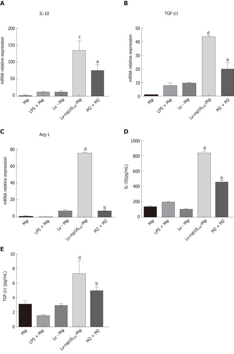
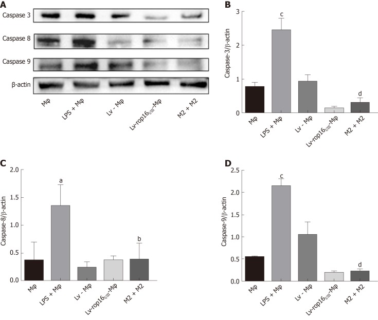
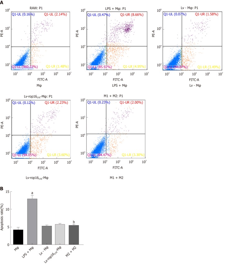
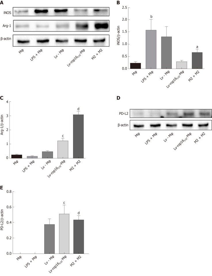
Methods: RAW264.7 macrophages stimulated by lipopolysaccharide (LPS) (M1 cells) were co-cultured with Caco-2 cells as an inflammatory model of IBD in vitro. The expression of ToxoROP16I/III was observed in RAW264.7 macrophages that were transfected with pEGFP-rop16 I/III. The phenotypes of M2 and M1 macrophage cells were assessed by quantitative real-time reverse transcriptase polymerase chain reaction and the expression of tumor necrosis factor (TNF)-α, interleukin (IL)-1β, IL-6, transforming growth factor (TGF)-β1, IL-10, inducible nitric oxide synthase (iNOS), and arginase-1 (Arg-1) was detected. The expression of iNOS, Arg-1, signal transducer and activator of transcription 3 (Stat3), p-Stat3, Stat6, p-Stat6, programmed death ligand-2 (PD-L2), caspase-3, -8, and -9 was analyzed by Western blotting, and Griess assays were performed to detect nitric oxide (NO). TNF-α, IL-1β, IL-6, TGF-β1, and IL-10 expression in the supernatants was detected by enzyme-linked immunosorbent assay, and Caco-2 cell apoptosis was determined by flow cytometry after mixing M1 cells with M2 cells in a Caco-2 cell co-culture system.

Results: M1 cells exhibited significantly increased production of iNOS, NO, TNF-α, IL-1β, and IL-6, while ToxoROP16I/III induced macrophage bias to M2 cells in vitro, showing increased expression of Arg-1, IL-10 and TGF-β1 and elevated production of p-Stat3 and p-Stat6. The mixed M1 and M2 cell culture induced by ToxoROP16I/III exhibited decreased production of NO and iNOS and upregulated expression of Arg-1 and PD-L2. Accordingly, Caco-2 cells became apoptotic, and apoptosis-associated proteins such as caspase-3, -8 and -9 were dampened during co-culture of M1 and M2 cells. Flow cytometry analysis showed that co-culture of M1 cells with Caco-2 cells facilitated the apoptosis of Caco-2 cells, but co-culture of M1 and M2 cells alleviated Caco-2 cell apoptosis.

Conclusion: ToxoROP16I/III-induced M2 macrophages inhibited apoptosis of Caco-2 cells caused by M1 macrophages. This finding may help gain a better understanding of the underlying mechanism and represent a promising therapeutic strategy for IBDs.

Keywords: Alternatively activated macrophages; Caco-2; Classically activated macrophages; Immunity; Inflammatory bowel disease; Toxoplasma ROP16I/III.


Conflict of interest statement

Conflict-of-interest statement: None of the authors has any conflicts of interest to declare.

Figures

Figure 1
Figure 1
Stable transfection of RAW264.7 cells with LV-rop16I/III recombinant lentivirus. A: Fluorescence microscopy was used to observe the expression of green fluorescent protein in macrophages, Lv-Mφ and Lv-rop16I/III-Mφ cells stably transfected with recombinant lentivirus. B: Macrophages, Lv-Mφ, and Lv-rop16I/III-Mφ stably-transfected cells were analyzed by Western blotting. C: Statistical analysis of protein expression in Lv-rop16I/III-Mφ cells relative to non-transfected macrophages and mock Lv-Mφ by Western blotting. aP < 0.001 vs Mφ. Mφ: Macrophages; LV-Mφ: Lentivirus transfer into macrophages; LV-rop16I/III-Mφ: Lentivirus-rop16I/III transfer into macrophages.
Figure 2
Figure 2
Lipopolysaccharide polarized to M1 cells. Protein was extracted from M1 cells at different time intervals over 24 h. iNOS expression was significantly increased at 6 h. iNOS: Inducible nitric oxide synthase.
Figure 3
Figure 3
The proinflammatory cytokines produced by M1, and mixed M1 and M2 cells. A-D: The mRNA expression levels of interleukin (IL)-6, IL-1β, tumour necrosis factor (TNF)-α, and iNOS were significantly increased in M1 cells by quantitative real-time reverse transcriptase polymerase chain reaction. Levels were normalized to those of β-actin. E-G: E-G: Expression of IL-6, IL-1β, and TNF-α was remarkably increased in the M1 cells in comparison with the control when examined by ELISA. D: The relative mRNA expression of iNOS was also increased in the M1 cell group. H: The nitric oxide (NO) was upregulated relative to normal macrophages as measured by the Griess method. The production of proinflammatory cytokines was markedly decreased in mixed M1 and M2 cells compared with M1 cells. The NO was measured by the Griess method. aP < 0.001 vs Mφ; bP < 0.001 vs lipopolysaccharide + Mφ. iNOS: Inducible nitric oxide synthase; NO: Nitric oxide; IL: Interleukin; LPS: Lipopolysaccharide; LV- Mφ: Lentivirus transfer into macrophages; LV-rop16I/III- Mφ: Lentivirus-rop16I/III transfer into macrophages.
Figure 4
Figure 4
Western blotting analysis for the detection of M1 and M2 cell signatures. A-C: LV-rop16I/III polarized macrophages to M2 cells via the phosphorylation of Stat3 and Stat6. The expression of p-Stat3 and p-Stat6 was significantly elevated in M2 cells relative to normal macrophages. D and E: The expression of PD-L2 (33 kDa) was obviously increased in M2 cells while Lv-Mφ was significantly increased in M1 cells relative to macrophages. F-H: The expression of Arg-1 was obviously increased in M2 cells while iNOS was significantly increased in M1 cells relative to macrophages. aP < 0.01 vs Lv-Mφ; bP < 0.001 vs Lv-Mφ; cP < 0.001 vs Mφ. Stat3: Signal transducer and activator of transcription 3; Stat6: Signal transducer and activator of transcription 6; p-Stat3: Phosphorylate signal transducer and activator of transcription 3; p-Stat6: Phosphorylate signal transducer and activator of transcription 6; iNOS: Inducible nitric oxide synthase; PD-L2: Programmed death ligand-2; Arg-1: Arginase-1; Mφ: Macrophages; LPS: Lipopolysaccharide; LV-Mφ: Lentivirus transfer into macrophages; LV-rop16I/III-Mφ: Lentivirus-rop16I/III transfer into macrophages.
Figure 5
Figure 5
Cytokine expression was detected in M2 eclls and mixed M1 and M2 cells. A-C: The relative mRNA expression of interleukin (IL)-10, transforming growth factor (TGF)-β1 and Arg-1 in M2 cells was markedly upregulated in Lv-rop16I/III-Mφ cells relative to LV-Mφ cells, while the production of cytokines was increased in mixed M1 and M2 cells relative to M1 cells. D and E: The culture supernatants were collected and analyzed by ELISA for IL-10 and TGF-β1, consistent with the result of relative mRNA. aP < 0.05 vs lipopolysaccharide (LPS) + Mφ; bP < 0.001 vs LPS + Mφ; cP < 0.01 vs Lv-Mφ, dP < 0.001 vs Lv-Mφ. Mφ: Macrophages; LV-Mφ: Lentivirus transfer into macrophages; LV-rop16I/III-Mφ: Lentivirus-rop16I/III transfer into macrophages.
Figure 6
Figure 6
Caco-2 cell apoptosis was restrained by M1 cells mixed with M2 cells. Lipopolysaccharide (LPS)-induced macrophages with the M1-like phenotype were co-cultured with Caco-2 cells. A-D: Western blotting indicated that the expression of caspase-3, caspase-8, and caspase-9 was significantly increased in M1 cells compared to normal macrophages. M1 cells and M2 cells were co-cultured with Caco-2 cells, the expression levels of apoptosis-associated proteins were significantly reduced compared to the M1 cell group. The above proteins were detected by Western blotting, and the data were analyzed by grey values. aP < 0.01 vs Mφ; bP < 0.01 vs LPS + Mφ; cP < 0.001 vs Mφ, dP < 0.001 vs LPS + Mφ. Mφ: Macrophages; LV-Mφ: Lentivirus transfer into macrophages; LV-rop16I/III-Mφ: Lentivirus-rop16I/III transfer into macrophages; LPS: Lipopolysaccharide.
Figure 7
Figure 7
M1 cells mixed with M2 cells lead to reduction of Caco-2 cell apoptosis in co-culture. A: M1 cells co-cultured with Caco-2 cells for 24 h induced higher Caco-2 cell apoptosis relative to normal macrophages, as detected by FCM. A and B: M1 cells mixed with M2 cells in the co-culture system resulted in a notable decrease in Caco-2 cell apoptosis M1 cells. aP < 0.001 vs Mφ; bP < 0.001 vs lipopolysaccharide + Mφ. Mφ: Macrophages; LPS: Lipopolysaccharide; LV- Mφ: Lentivirus transfer into macrophages; LV-rop16I/III - Mφ: Lentivirus-rop16I/III transfer into macrophages; LPS: Lipopolysaccharide.
Figure 8
Figure 8
M2 cells reduced iNOS expression in M1 inflammatory macrophages. LV-rop16I/III-induced M2-like macrophages restricted the production of iNOS in M1 cells. A and B: The iNOS level was downregulated in mixed M1 and M2 cells relative to that in M1 cells. A, C-E: Arg-1 and PD-L2 expression was higher in M2 cells than in Lv-Mφ cells. The above proteins were detected by Western blotting, and the data were analyzed by grey values. aP < 0.05 vs lipopolysaccharide (LPS) + Mφ; bP < 0.01 vs Mφ; cP < 0.001 vs Lv-Mφ, dP < 0.001 vs LPS + Mφ. iNOS: Inducible nitric oxide synthase; PD-L2: Programmed death ligand-2; Arg-1: Arginase-1; Mφ: Macrophages; LPS: Lipopolysaccharide; LV-Mφ: Lentivirus transfer into macrophages; LV-rop16I/III-Mφ: Lentivirus-rop16I/III transfer into macrophages.

Similar articles

Cited by

References

    1. Corridoni D, Arseneau KO, Cominelli F. Inflammatory bowel disease. Immunol Lett. 2014;161:231–235. - PMC - PubMed
    1. Markowitz J, Grancher K, Kohn N, Daum F. Immunomodulatory therapy for pediatric inflammatory bowel disease: changing patterns of use, 1990-2000. Am J Gastroenterol. 2002;97:928–932. - PubMed
    1. Ananthakrishnan AN. Epidemiology and risk factors for IBD. Nat Rev Gastroenterol Hepatol. 2015;12:205–217. - PubMed
    1. Cader MZ, Kaser A. Recent advances in inflammatory bowel disease: mucosal immune cells in intestinal inflammation. Gut. 2013;62:1653–1664. - PubMed
    1. Kim JK, Jun JG. Ailanthoidol suppresses lipopolysaccharide-stimulated inflammatory reactions in RAW264.7 cells and endotoxin shock in mice. J Cell Biochem. 2011;112:3816–3823. - PubMed

MeSH terms