Review
. 2019 Feb 1;8(2):77-89.
doi: 10.1089/wound.2018.0828. Epub 2019 Feb 13.

Animal Models for Studies of Keloid Scarring

Affiliations
Review

Animal Models for Studies of Keloid Scarring

Dorothy M Supp. Adv Wound Care (New Rochelle). .

Abstract

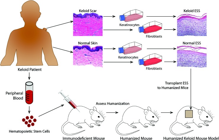
Significance: Keloid scarring is a disfiguring fibroproliferative disorder that can significantly impair the quality of life in affected individuals. The mechanisms that initiate keloid scarring are incompletely understood, and keloids remain one of the most challenging skin conditions to treat. Keloids are unique to humans; thus, the lack of adequate animal models has hindered research efforts aimed at prevention and effective therapeutic intervention. Recent Advances: In the absence of a suitable animal model, keloid researchers often rely on studying excised keloid scar tissue and keloid-derived cultured cells. Recently, in vivo models have been described that involve transplantation to mice of reconstructed skin containing keloid-derived fibroblasts and/or keratinocytes. These mouse-human hybrid animal models display some similarities with keloids and may enable investigation of novel therapies, although no model yet recapitulates all the features of human keloid scarring. Critical Issues: Differences in skin physiology and modes of healing contribute to challenges in modeling keloids in laboratory animals. Furthermore, recent studies suggest that cells of the immune system contribute to keloid pathology. The need to use immunodeficient hosts for transplanted human keloid cells in recently described animal models precludes studying the role of the immune system in keloid scarring. Future Directions: Future animal models may take advantage of humanized mice with immune systems reconstituted using human immune cells. Such models, when combined with grafted tissues prepared using keloid-derived cells, might enable investigation of complex interactions between systemic and local factors that combine to promote keloid scar formation and may aid in the development of novel therapies.

Keywords: animal model; extracellular matrix; fibrosis; keloid; scar; wound healing.


Figures

None
Dorothy M. Supp, PhD
<b>Figure 1.</b>
Figure 1.
Keloid scar development. Shown are photos of the same patient illustrating the rapid development of keloid lesions over time after skin injury. The patient, an African American male who sustained a 15% total body surface area burn at 15 years of age, developed widespread keloid lesions in both grafted and ungrafted burn wounds, as well as donor sites used for autograft harvesting. (AC) Images of patient's left shoulder, showing a healed partial-thickness burn wound that was not grafted. Note the rapid development of large keloid with typical bulging appearance. (DF) Images of patient's right shoulder; this deeper burn wound was treated with split-thickness skin autograft. Note the development of keloid scarring around the skin graft and within the grafted area where wounds appear to have occurred. Photographs were taken at PBD 72 (A, D), PBD 114 (B, E), and PBD 332 (C, F). PBD, postburn day. Color images are available online.
<b>Figure 2.</b>
Figure 2.
Normal and keloid ESS in vivo. (AD) Shown are histological sections of normal (A, B) and keloid (C, D) ESS 12 weeks after transplantation to mice. Sections in the top panels (A, C) were stained with Tango stain, similar to hematoxylin; bottom panels (B, D) show Masson's trichrome-stained sections. Note the densely packed disorganized collagen fibers in the keloid ESS. Scale bars for all sections, 200 μm. (E) Quantitation of the total thickness of the grafted tissue demonstrated that ESS prepared with keloid cells were significantly thicker than ESS prepared using normal cells. ESS, engineered skin substitutes. Color images are available online.
<b>Figure 3.</b>
Figure 3.
Model for the development of bulging keloid scars. Analysis of deep versus superficial keloid fibroblasts in engineered skin grafted to mice suggested that deep fibroblasts contribute to graft thickening, whereas superficial fibroblasts induce a spreading phenotype. Other studies described the migratory phenotype of keloid keratinocytes., Together, these observations contributed to the model illustrated here. Shown at the left is a schematic diagram of a cross-section of skin following a wound. During wound healing and over time, fibroblasts proliferate, migrate, and deposit ECM to form granulation tissue over which keratinocytes migrate to close the wound. For reasons that have yet to be identified, cells in scars that progress to keloids fail to respond to “stop” signals, and proliferation, ECM production, and migration continue unchecked. Continued production of ECM in fibroblasts in the deep dermis contributes to thickening of the lower dermis, while fibroblasts in the upper dermis exhibit a spreading phenotype, causing an increase in area. With increasing time after injury, the combination of deep dermal thickening and superficial spreading results in a bulging phenotype. Figure adapted with permission from Supp et al. ECM, extracellular matrix. Color images are available online.
<b>Figure 4.</b>
Figure 4.
Use of humanized mice to study keloid scar development. This schematic diagram illustrates the potential use of humanized mice as hosts for grafting of ESS prepared using keloid-derived or normal skin-derived fibroblasts and keratinocytes. Humanized mice are prepared by injection of CD34+ hematopoietic stem cells into severely immunodeficient mice (see text for details). Resulting mice harbor immune systems reconstituted by human cells, enabling studies of the human immune response in a mouse experimental model. Grafting of ESS containing keloid-derived cells to humanized mice can permit investigation of the role of the immune system in keloid scar development, which is currently not possible using standard immunodeficient mouse hosts. ESS containing normal cells can be compared with ESS containing keloid-derived cells to determine the relative contribution(s) of skin cells and immune cells in keloid pathology. This diagram shows images of ESS prepared using primary keratinocytes and fibroblasts cultured from keloid scar and normal skin. Melanocytes and microvascular endothelial cells have also been used in preparation of ESS; thus, this model can be used to study the relative roles of numerous different cell types in keloid pathology. Comparison of mice humanized with keloid patient-derived hematopoietic stem cells versus normal donor stem cells can be used to identify specific components of the immune system involved in keloid development. Currently, isolation of sufficient numbers of hematopoietic stem cells from peripheral blood is an obstacle to implementation of such a model, but future developments aimed at expansion of this population and improved methods for stem cell recovery are expected to enable such studies in the near future. Color images are available online.

Similar articles

Cited by

References

    1. Van Loey NEE, Van Son MJM. Psychopathology and psychological problems in patients with burn scars. Am J Clin Dermatol 2003;4:245–272 - PubMed
    1. Bock O, Schmid-Ott G, Malewski P, Mrowietz U. Quality of life of patients with keloid and hypertrophic scarring. Arch Dermatol Res 2006;297:433–438 - PubMed
    1. Balci DD, Inandi T, Dogramaci CA, Celik E. DLQI scores in patients with keloids and hypertrophic scars: a prospective case control study. J Dtsch Dermatol Ges 2009;7:688–692 - PubMed
    1. Furtado F, Hochman B, Ferrara SF, et al. . What factors affect the quality of life of patients with keloids? Rev Assoc Med Bras 2009;55:700–704 - PubMed
    1. Chike-Obi CJ, Cole PD, Brissett AE. Keloids: pathogenesis, clinical features, and management. Semin Plast Surg 2009;23:178–184 - PMC - PubMed

LinkOut - more resources