

Treatment of Periorbital and Palpebral Arteriovenous Malformations
- PMID: 31832275
- PMCID: PMC6906756
- DOI: 10.1089/wound.2018.0846
Treatment of Periorbital and Palpebral Arteriovenous Malformations
Abstract
Objectives: To clarify clinically challenging palpebral arteriovenous malformations (AVMs) and to propose a novel therapeutic modality, we developed a multi-disciplinary approach for the management of AVMs with ulcer. Approach: First, the central retinal artery was secured with embolization by the transophthalmic arterial, a terminal branch of the internal carotid artery (ICA), and then, the branches of the external carotid artery (ECA) were embolized to cause a response in the AVM vasculature followed by sclerotherapy and surgery. Results: Over a 3-year follow-up of palpebral and periorbital AVMs in four females and one male 20 to 50 years of age with a mean age of 38 years, complete remission of the lesions were seen with no major complication, such as blindness, ptosis, or cerebral infarction, with functionally sound and esthetically acceptable results, with no recurrence or worsening even with one case of ulceration postembolization. Innovation: Planned treatment of palpebral and periorbital AVMs, which have been often left untreated because of their complex vasculature and a risk of total blindness due to occlusion of the central retinal artery. A "wait-and-watch" approach is frequently taken. It is important to secure the periphery to the bifurcation of the central retinal artery of the ICA, and then, embolization through the ECA results in complete remission of the lesion, followed by sclerotherapy and surgery, which are successful both in terms of function and esthetics. Conclusion: First, securing the central retinal artery leads to safer and complete resolution of palpebral and periorbital AVMs; wounding or therapeutic complications such as skin necrosis may be seen, but this approach results in complete remission in 3 years with no major complications.
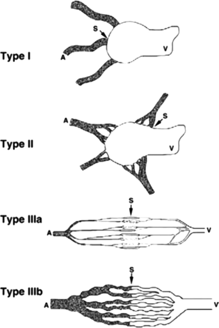
Keywords: Schobinger staging; angiographic diagram; central retinal artery; external carotid artery; following sclerotherapy and surgery; transophthalmic arterial embolization.
Copyright 2019, Mary Ann Liebert, Inc., publishers.
Figures


References
-
- Kohout MP, Hansen M, Pribaz JJ, Mulliken JB. Arteriovenous malformations of the head and neck: natural history and management. Plast Reconstr Surg 1998;102:643–654 - PubMed
-
- Enjolras O, Logeart I, Gelbert F, et al. . Arteriovenous malformations: a study of 200 cases. Ann Dermatol Venereol 2000;127:17–22 - PubMed
-
- Elluru RG, Azizkhan RG. Cervicofacial vascular anomalies. II. Vascular malformations. Semin Pediatr Surg 2006;15:133–139 - PubMed
-
- Liu AS, Mulliken JB, Zurakowski D, Fishman SJ, Greene AK. Extracranial arteriovenous malformations: natural progression and recurrence after treatment. Plast Reconstr Surg 2010;125:1185–1194 - PubMed
-
- Cahill AM, Nijs EL. Pediatric vascular malformations: pathophysiology, diagnosis, and the role of interventional radiology. Cardiovasc Intervent Radiol 2011;34:691–704 - PubMed
Publication types
LinkOut - more resources
Full Text Sources
Miscellaneous
