Review
. 2019 Nov;8(6):691-698.
doi: 10.21037/acs.2019.11.05.

The "UFO" procedure

Affiliations
Review

The "UFO" procedure

Martin Misfeld et al. Ann Cardiothorac Surg. 2019 Nov.

Abstract

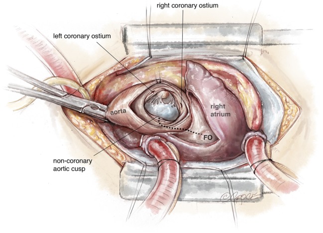
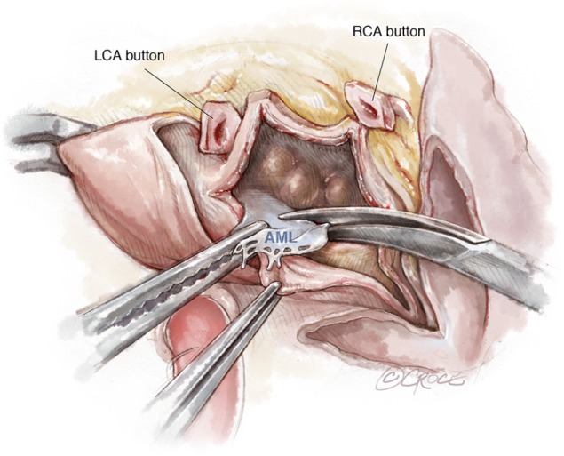
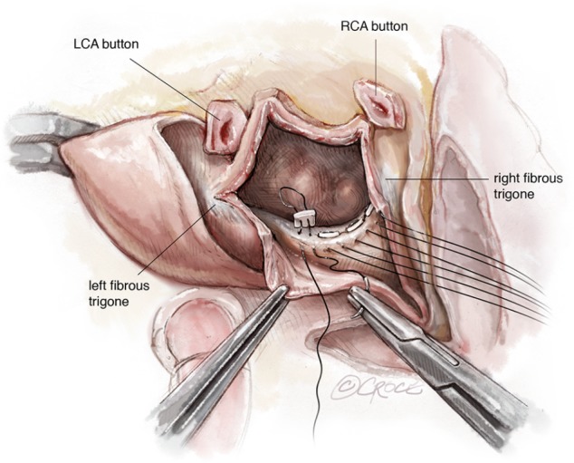
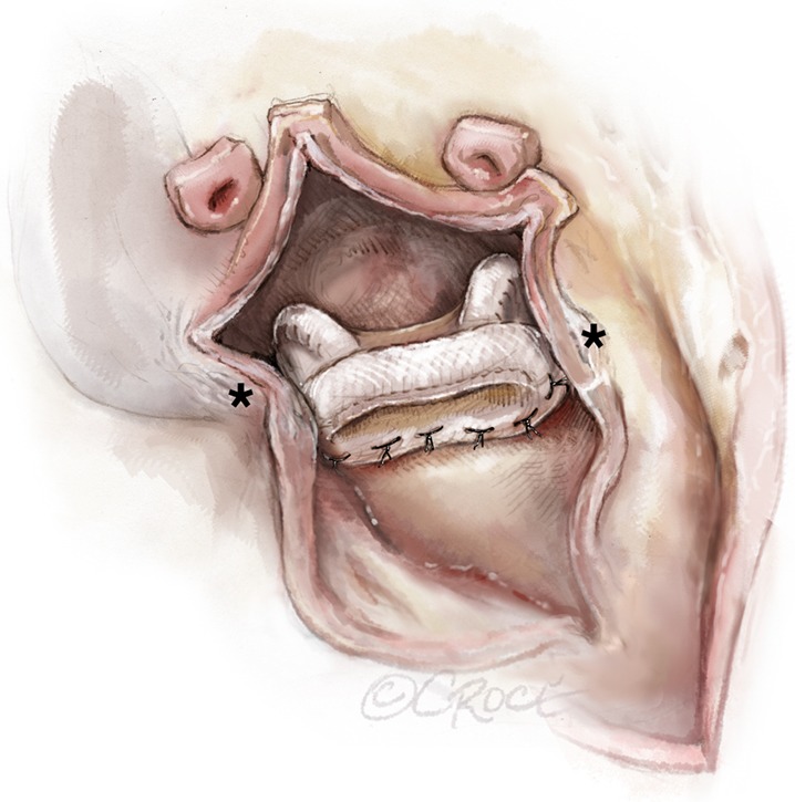
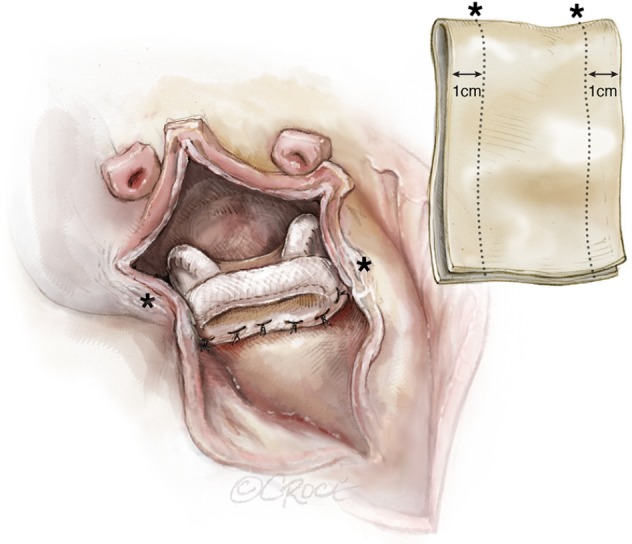
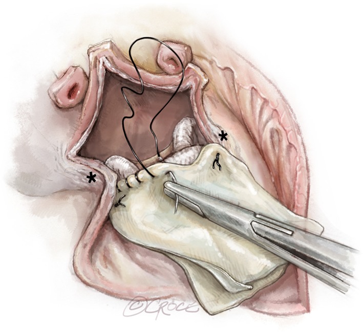
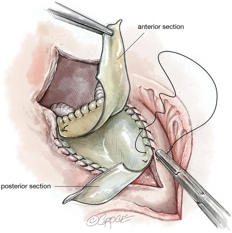
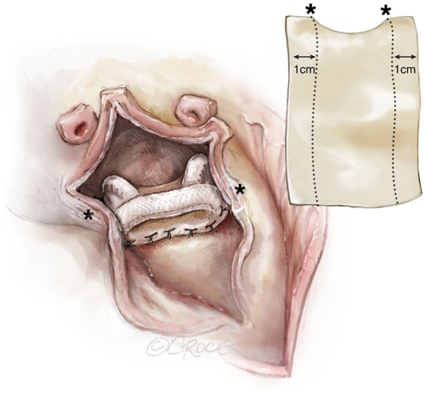
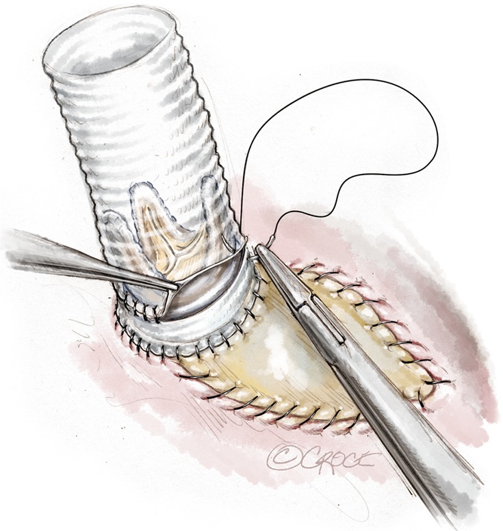
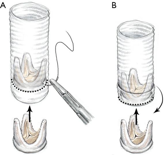
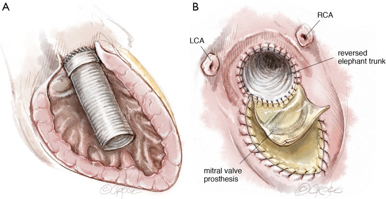
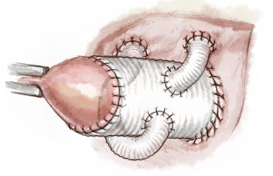
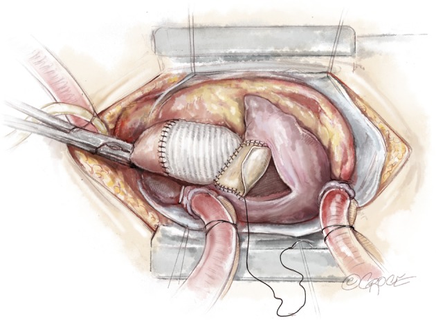
The term "UFO" is not a medical term, but helps emphasize the extremely high degree of complexity of a surgical repair that is akin to someone observing an unidentified flying object. It involves replacement of the mitral and aortic valves with reconstruction of the intervalvular fibrous body (IVFB). Specific pathologies that render this operation necessary usually involve the IVFB, which is located between the aortic and mitral valves and constitutes a major portion of the fibrous skeleton of the heart. Patients that most often require such an operation are those with extensive aortic and mitral valve endocarditis with perivalvular extension into the IVFB. Other infrequent situations such as severe aortic and mitral annular calcification involving the IVFB, double valve replacement in patients with extremely small aortic and mitral annuli or double valve reoperations in which no IVFB is available following excision of both valves, necessitating the UFO procedure. The basic surgical principle has been first described as early as 1980. Depending on the extent of excised tissue due to the underlying disease, modifications and additional complex repair techniques have to be adopted. It is of utmost importance to have adequate visibility and exposure. There are certain important structures, which are at a risk of either injury or neglect, that can result in development of life-threatening complications during this operation, which a surgeon should be aware of. A step by step description of the "UFO" procedure can help guide the surgeon to perform this operation safely and efficiently. Although clinical complications are high, they are often related to the underlying disease and not specifically to the procedure itself, if performed perfectly.

Keywords: Valve surgery; endocarditis; fibrous body.


Conflict of interest statement

Conflicts of Interest: The authors have no conflicts of interest to declare.

Figures

Figure 1
Figure 1
An oblique aortic incision extending into the non-coronary sinus. Incision into the lateral wall of the right atrium extending across the appendage into its medial wall towards the aortic root. Hatched line: incision of the atrial septum, beginning from the fossa ovalis extending into the roof of the left atrium towards the midpoint of the non-coronary sinus. FO, fossa ovalis.
Figure 2
Figure 2
The AML is resected. Coronary artery buttons are prepared. The aortic root, left atrium and left ventricle are inspected. LCA, left coronary artery; RCA, right coronary artery; AML, anterior mitral leaflet.
Figure 3
Figure 3
View as in Figure 2, but with pledgeted valve sutures in place in the posterior mitral valve annulus from the lateral to medial fibrous trigones. LCA, left coronary artery; RCA, right coronary artery.
Figure 4
Figure 4
A mitral bioprosthesis fixed posteriorly from the left to the right fibrous trigones (marked with *). Note the position of the struts, avoiding obstruction of the left ventricular outflow tract.
Figure 5
Figure 5
Trimming and sizing of the double patch. The marked distance on the patch corresponds to the distance between the trigones (*). A 1 cm margin is kept for attachment to the left atrium.
Figure 6
Figure 6
Double patch technique. The folded margin of the patch is sutured to the anterior sewing cuff of the mitral valve prosthesis. Reinforcement sutures are placed at the trigones.
Figure 7
Figure 7
Double patch technique. The posterior tongue of the patch is used to reconstruct the left atrial roof. The anterior tongue is used to reconstruct parts of the aortic root or for anchoring the aortic valve conduit.
Figure 8
Figure 8
Single patch technique for reconstruction of the left atrial roof. The distance between the marks (*) corresponds to the distance between the trigones. The patch may be trimmed to replicate the curvature of the prosthesis.
Figure 9
Figure 9
A skirt of around 1–1.5 cm is kept under the aortic prosthetic valve and used for anchoring the conduit into the LVOT. This length can be variable, depending, how deep the conduit has to be inserted into the LVOT and the level of the coronary buttons. LVOT, left ventricular outflow tract.
Figure 10
Figure 10
Composition of a “bio-Bentall” conduit. An aortic valve prosthesis is inserted into the Dacron graft at the desired level and secured by suturing it to the graft from outside (A); alternatively, the Dacron graft is folded on itself and the prosthesis is directly sutured to the folded margin of the graft. Thereafter, the graft is straightened by pulling the folded part over the valve, thus forming a skirt under it (B).
Figure 11
Figure 11
Anchoring of a vascular graft in the LVOT. Dacron graft is inverted into the left ventricle (A); the folded margin of the Dacron graft is then anchored to the LVOT with a double layer of sutures (B). LCA, left coronary artery; RCA, right coronary artery; LVOT, left ventricular outflow tract.
Figure 12
Figure 12
Modified Cabrol procedure: Single Dacron grafts (size, 6–8 mm) are anastomosed to each coronary ostium and reimplanted into the ascending aortic graft.
Figure 13
Figure 13
Reconstruction of the right atrium using a pericardial patch.

References

    1. Manouguian S. Erweiterungsplastik des hypo-plastischen Aortenklappenringes: Beschreibung einer neuen Operationsmethode: eine experimentelle Studie. Thorac Cardiov Surg 1976;24:418-23. 10.1055/s-0028-1095957 - DOI - PubMed
    1. Manouguian S, Kirchhoff PG. Patch enlargement of the aortic and the mitral valve rings with aortic-mitral double-valve replacement. Ann Thorac Surg 1980;30:396-9. 10.1016/S0003-4975(10)61281-7 - DOI - PubMed
    1. David TE, Kuo J, Armstrong S. Aortic and mitral valve replacement with reconstruction of the intervalvular fibrous body. J Thorac Cardiovasc Surg 1997;114:766-71; discussion 771-2. 10.1016/S0022-5223(97)70080-1 - DOI - PubMed
    1. Davierwala PM, Binner C, Subramanian S, et al. Double valve replacement and reconstruction of the intervalvular fibrous body in patients with active infective endocarditis. Eur J Cardiothorac Surg 2014;45:146-52. 10.1093/ejcts/ezt226 - DOI - PubMed
    1. Forteza A, Centeno J, Ospina V, et al. Outcomes in aortic and mitral valve replacement with intervalvular fibrous body reconstruction. Ann Thorac Surg 2015;99:838-45. 10.1016/j.athoracsur.2014.09.052 - DOI - PubMed