The Role of digital subtraction angiography in the ventricular spot sign on the computed tomography angiography
- PMID: 31832384
- PMCID: PMC6901812
- DOI: 10.7461/jcen.2019.21.1.24
The Role of digital subtraction angiography in the ventricular spot sign on the computed tomography angiography
Abstract
Objective: The spot sign on computed tomography angiography is little known about the relationship between the spot sign and the results of cerebral angiography We retrospectively analyzed the spot sign, digital subtraction angiography results, and other factors.
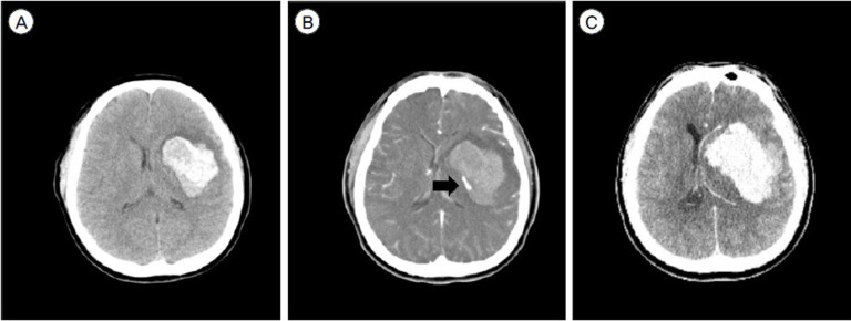
Material and methods: From December 2009 to May 2014, DSA was performed in 52 ICH patients with non-specific location or abnormalities on CTA findings. 26 of those patients, whose initial CTA showed the spot sign, were analyzed. Two groups, one with the spot sign in the ventricle (Group A) and others with the spot sign in another location (Group B) were statistically compared.
Results: The mean age of the study subjects was 46.9 years (range, 15 to 80 years) and the percentage of males was 53.8%. Thirteen of 26 patients had ICH without intraventricular hemorrhage, and 6 patients had co-existing IVH. In 17 cases, the DSA results were negative. Seven patients were diagnosed with pseudoaneurysms, and two cases showed developmental venous anomalies. Group A consisted of the 8 patients (30.8%) who showed the spot sign in a ventricle. The number of pseudoaneurysms was statistically significantly higher in Group A than in Group B (71.4% versus 28.6%; OR, 13.3; 95% CI, 1.7-103.8 P = 0.014). All three patients who underwent endovascular treatment were members of Group A (P = 0.022), whereas most (92.3%) of those in Group B underwent surgical evacuation. (P = 0.030).
Conclusion: When CTA shows the spot sign in a ventricle, it is a clue that an existing underlying vascular lesion requires endovascular treatment.
Keywords: Cerebral angiography; Cerebral hemorrhage; computed tomography angiography; spot sign.
© 2019 Journal of Cerebrovascular and Endovascular Neurosurgery.
Figures


References
-
- Brzozowski K, Frankowska E, Piasecki P, Ziecina P, Zukowski P, Boguslawska-Walecka R. The use of routine imaging data in diagnosis of cerebral pseudoaneurysm prior to angiography. Eur J Radiol. 2011 Dec;80(3):e401–e409. - PubMed
-
- Ciceri EF, Regna-Gladin C, Erbetta A, Chiapparini L, Nappini S, Savoiardo M, et al. Iatrogenic intracranial pseudoaneurysms: neuroradiological and therapeutical considerations, including endovascular options. Neurol Sci. 2006 Nov;27(5):317–322. - PubMed
-
- Davis SM, Broderick J, Hennerici M, Brun NC, Diringer MN, Mayer SA, et al. Hematoma growth is a determinant of mortality and poor outcome after intracerebral hemorrhage. Neurology. 2006 Apr;66(8):1175–1181. - PubMed
-
- de Andrade AF, Figueiredo EG, Caldas JG, Paiva WS, De Amorim RL, Puglia P, et al. Intracranial vascular lesions associated with small epidural hematomas. Neurosurgery. 2008 Feb;62(2):416–420. discussion 20–1. - PubMed
-
- Demchuk AM, Dowlatshahi D, Rodriguez-Luna D, Molina CA, Blas YS, Dzialowski I, et al. Prediction of haematoma growth and outcome in patients with intracerebral haemorrhage using the CT-angiography spot sign (PREDICT): a prospective observational study. The Lancet Neurology. 2012;11(4):307–314. - PubMed