

Dual time point [18F]FLT-PET for differentiating proliferating tissues vs non-proliferating tissues
- PMID: 31832803
- PMCID: PMC6908533
- DOI: 10.1186/s13550-019-0579-5
Dual time point [18F]FLT-PET for differentiating proliferating tissues vs non-proliferating tissues
Abstract
Purpose: For differentiating tumor from inflammation and normal tissues, fluorodeoxyglucose ([18F]FDG) dual time point PET could be helpful. Albeit [18F]FLT is more specific for tumors than [18F]FDG; we explored the role of dual time point [18F]FLT-PET for discriminating benign from malignant tissues.
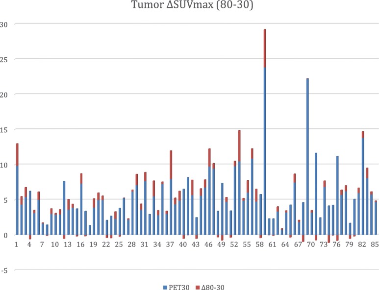
Methods: Before any treatment, 85 womens with de novo unifocal breast cancer underwent three PET acquisitions at 33.94 ± 8.01 min (PET30), 61.45 ± 8.30 min (PET60), and 81.06 ± 12.12 min (PET80) after [18F]FLT injection. Semiquantitative analyses of [18F]FLT uptake (SUV) were carried out on tumors, liver, bone marrow (4th thoracic vertebra (T4) and humeral head), descending thoracic aorta, muscle (deltoid), and contralateral normal breast. Repeated measures ANOVA tests and Tukey's posttests were used to compare SUVmax of each site at the three time points.
Results: There was a significant increase in SUVmax over time for breast lesions (5.58 ± 3.80; 5.97 ± 4.56; 6.19 ± 4.42; p < 0.0001) (m ± SD for PET30, PET60, and PET80, respectively), and bone marrow (for T4, 8.21 ± 3.17, 9.64 ± 3.66, 10.85 ± 3.63, p < 0.0001; for humeral head, 3.36 ± 1.79, 3.87 ± 1.89, 4.39 ± 2.00, p < 0.0001). A significant decrease in SUVmax over time was observed for liver (6.79 ± 2.03; 6.24 ± 1.99; 5.57 ± 1.74; p < 0.0001), muscle (0.95 ± 0.28; 0.93 ± 0.29; 0.86 ± 0.20; p < 0.027), and aorta (1.18 ± 0.34; 1.01 ± 0.32; 0.97 ± 0.30; p < 0.0001). No significant difference was observed for SUVmax in contralateral breast (0.8364 ± 0.40; 0.78 ± 0.38; 0.80 ± 0.35).
Conclusion: [18F]FLT-SUVmax increased between 30 and 80 min only in proliferating tissues. This could be helpful for discriminating between residual tumor and scar tissue.
Keywords: Breast cancer; FLT; Fluorothymidine; Positron emission tomography.
Conflict of interest statement
The authors declare that they have no competing interests
Figures
References
-
- Gallagher BM, Ansari A, Atkins H, Casella V, Christman DR, Fowler JS, et al. Radiopharmaceuticals XXVII. 18F-labeled 2-deoxy-2-fluoro-d-glucose as a radiopharmaceutical for measuring regional myocardial glucose metabolism in vivo: tissue distribution and imaging studies in animals. J Nucl Med. 1977;18:990–996. - PubMed
-
- Romain S, Martin P-M, Klijn JGM, van Putten WLJ, Look MP, Guirou O, et al. DNA-synthesis enzyme activity: a biological tool useful for predicting anti-metabolic drug sensitivity in breast cancer? International Journal of Cancer. Wiley-Blackwell. 1997;74:156–161. - PubMed
Grants and funding
LinkOut - more resources
Full Text Sources
