Review
. 2020 Feb 3;39(3):e102309.
doi: 10.15252/embj.2019102309. Epub 2019 Dec 12.

Molecular and physiological consequences of faulty eukaryotic ribonucleotide excision repair

Affiliations
Review

Molecular and physiological consequences of faulty eukaryotic ribonucleotide excision repair

Vanessa Kellner et al. EMBO J. .

Abstract

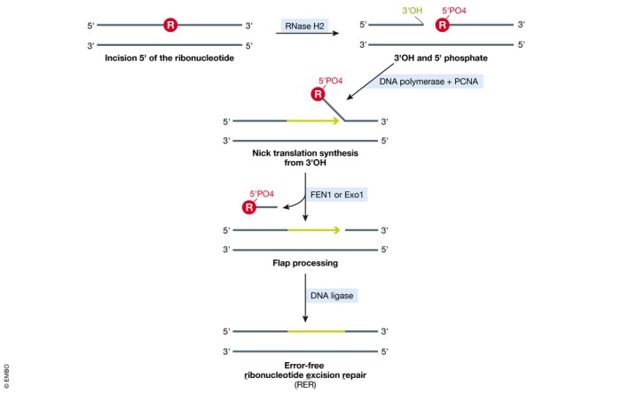
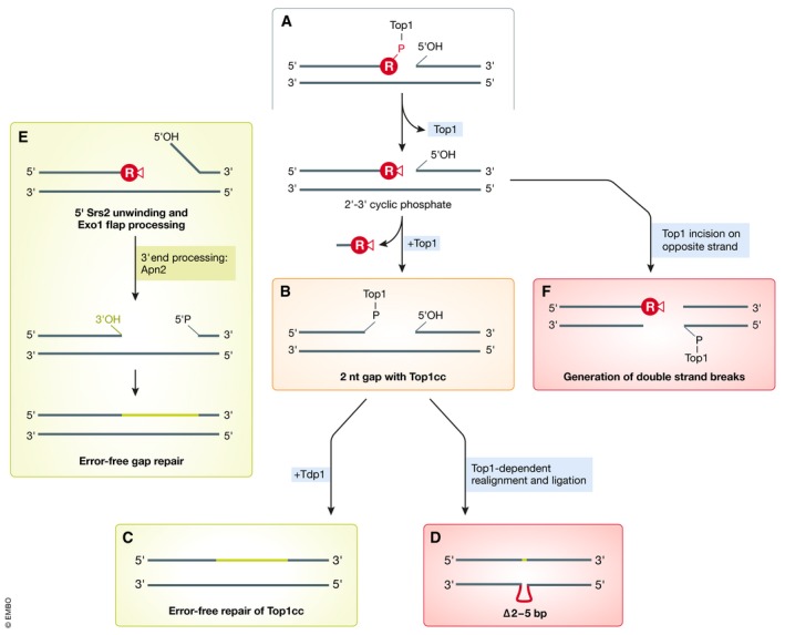
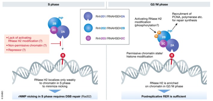
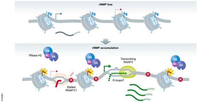
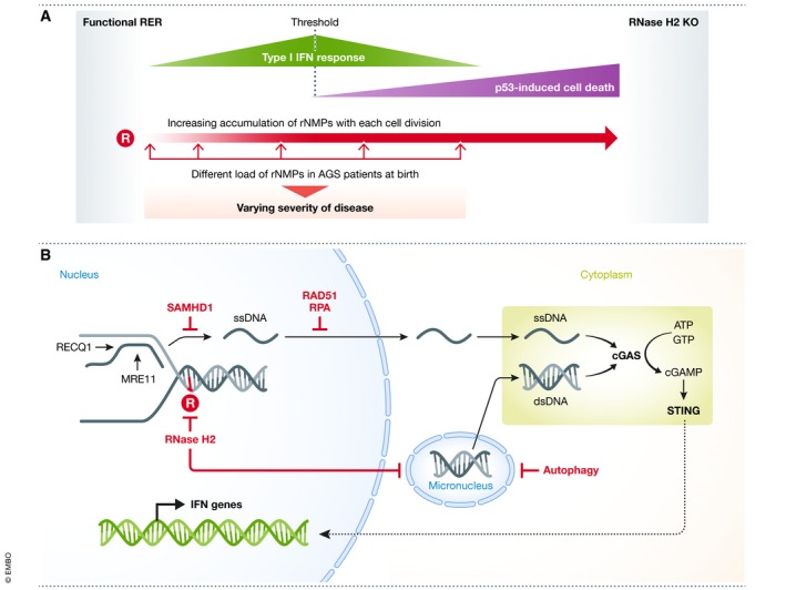
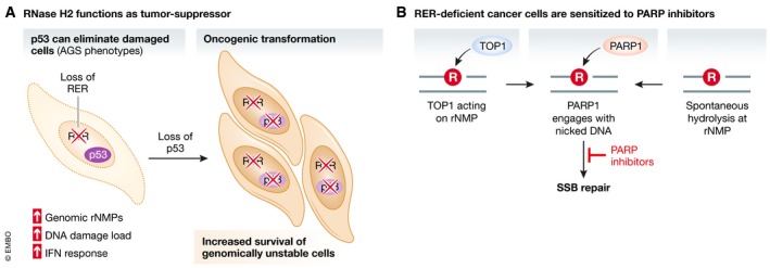
The duplication of the eukaryotic genome is an intricate process that has to be tightly safe-guarded. One of the most frequently occurring errors during DNA synthesis is the mis-insertion of a ribonucleotide instead of a deoxyribonucleotide. Ribonucleotide excision repair (RER) is initiated by RNase H2 and results in error-free removal of such mis-incorporated ribonucleotides. If left unrepaired, DNA-embedded ribonucleotides result in a variety of alterations within chromosomal DNA, which ultimately lead to genome instability. Here, we review how genomic ribonucleotides lead to chromosomal aberrations and discuss how the tight regulation of RER timing may be important for preventing unwanted DNA damage. We describe the structural impact of unrepaired ribonucleotides on DNA and chromatin and comment on the potential consequences for cellular fitness. In the context of the molecular mechanisms associated with faulty RER, we have placed an emphasis on how and why increased levels of genomic ribonucleotides are associated with severe autoimmune syndromes, neuropathology, and cancer. In addition, we discuss therapeutic directions that could be followed for pathologies associated with defective removal of ribonucleotides from double-stranded DNA.

Keywords: DNA repair; RNA-DNA hybrid; RNase H2; ribonucleotide excision repair; topoisomerase 1.


Conflict of interest statement

The authors declare that they have no conflict of interest.

Figures

Figure 1
Figure 1. Overview of ribonucleotide excision repair (RER)
RNase H2 initiates RER by incising the DNA backbone at the rNMP (R in red circle). Nick translation DNA synthesis from the newly created 3′OH followed by FEN1/Exo1‐mediated flap processing and subsequent DNA ligation can efficiently repair the incised DNA, resulting in removal of the rNMP.
Figure 2
Figure 2. Topoisomerase 1 as backup for RER in rNMP removal from the genome
(A) In the absence of functional RNase H2, Top1 can act on accumulating rNMPs. Different outcomes have been characterized in budding yeast (see text for detailed descriptions), resulting either in error‐free repair or in repair that causes mutations or potentially lethal double‐strand breaks. (B–D) Secondary Top1‐mediated incision two basepairs upstream releases an rNMP‐dNMP dinucleotide and creates a Top1‐linked gap (B) that can be processed in an error‐free manner via Tdp1 (C), or in an error‐prone manner caused by Top1 realignment and religation (D). (E) Error‐free gap repair based on subsequent activities of Srs2 helicase, Exo1 exonuclease, and Apn2 abasic endonuclease, which prevent erroneous religation. (F) Secondary Top1 incision on the opposite strand creates DNA double‐strand breaks that require repair by homologous recombination.
Figure 3
Figure 3. Regulation of RER through RNase H2
RNase H2 chromatin localization gradually increases throughout S phase but its activity may be kept in check to prevent creation of nicks during replication, where they would be converted into one‐ended DSBs by oncoming replication forks. Different regulatory mechanisms (in red) might account for RNase H2 inactivity during S phase. In G2/M, RNase H2 actively processes rNMPs to achieve successful RER.
Figure 4
Figure 4. Possible chromatin alterations associated with rNMPs
The accumulation or insertion of rNMPs (R in red circle) may affect the local chromatin environment by altering histone modifications on nucleosomes (flags) or even nucleosome stability and histone occupancy. These local chromatin changes may be important to allow RER by RNase H2. Such chromatin alterations may also lead to more open chromatin, increasing the expression of lowly expressed genes and thus enhancing formation of R‐loops. RNA polymerase stalling at persistent rNMPs may further impact on transcription processes.
Figure 5
Figure 5. Impact of AGS mutations and IFN gene activation in disease
(A) With increasing rNMP load in the genome, IFN‐regulated genes are upregulated. When rNMP levels cross a threshold, p53‐dependent cell death leads to a decreased IFN response but an enhanced phenotype. Different RNase H2 mutations identified in AGS patients could result in different initial rNMP levels at birth and explain the observed differences in disease severity between patients. (B) Release of nuclear DNA into the cytoplasm triggers activation of cGAS, which in turn activates STING that subsequently induces IFN genes. SAMHD1 prevents ssDNA release from stalled replication forks processed by RECQ1 and MRE11. RAD51 and RPA bind to and retain ssDNA in the nucleus. RNase H2 removes rNMPs (R in red circle) from DNA and thereby prevents formation of micronuclei. Autophagy can clear micronuclei, preventing them from rupture.
Figure 6
Figure 6. Tumor suppressor functions of RNase H2 and RER
(A) Loss of RER leads to increased genomic rNMP accumulation and the indicated consequences, but damaged cells are eliminated as long as p53 is present. In the nervous system, cell elimination likely leads to the neurodegenerative phenotypes associated with AGS. Genome instability upon p53 loss can lead to oncogenic rearrangements and cancer development. (B) rNMPs that accumulate in the absence of RER are either cleaved by TOP1 or hydrolyzed spontaneously, creating single‐strand breaks (SSB). These nicks recruit PARP, sensitizing cells to PARP inhibitors that will create toxic PARP‐trapping lesions.

Similar articles

Cited by

References

    1. Aden K, Bartsch K, Dahl J, Reijns MAM, Esser D, Sheibani‐Tezerji R, Sinha A, Wottawa F, Ito G, Mishra N et al (2019) Epithelial RNase H2 maintains genome integrity and prevents intestinal tumorigenesis in mice. Gastroenterology 156: 145–159.e19 - PMC - PubMed
    1. Ahel I, Rass U, El‐Khamisy SF, Katyal S, Clements PM, McKinnon PJ, Caldecott KW, West SC (2006) The neurodegenerative disease protein aprataxin resolves abortive DNA ligation intermediates. Nature 443: 713–716 - PubMed
    1. Akwa Y, Hassett DE, Eloranta M‐L, Sandberg K, Masliah E, Powell H, Whitton JL, Bloom FE, Campbell IL (1998) Transgenic expression of IFN‐α in the central nervous system of mice protects against lethal neurotropic viral infection but induces inflammation and neurodegeneration. J Immunol 161: 5016–5026 - PubMed
    1. Allen‐Soltero S, Martinez SL, Putnam CD, Kolodner RD (2014) A Saccharomyces cerevisiae RNase H2 interaction network functions to suppress genome instability. Mol Cell Biol 34: 1521–1534 - PMC - PubMed
    1. Arana ME, Kerns RT, Wharey L, Gerrish KE, Bushel PR, Kunkel TA (2012) Transcriptional responses to loss of RNase H2 in Saccharomyces cerevisiae . DNA Repair (Amst) 11: 933–941 - PMC - PubMed

Publication types