

Sevoflurane postconditioning improves spatial learning and memory ability involving mitochondrial permeability transition pore in hemorrhagic shock and resuscitation rats
- PMID: 31833229
- PMCID: PMC6955830
- DOI: 10.1002/brb3.1501
Sevoflurane postconditioning improves spatial learning and memory ability involving mitochondrial permeability transition pore in hemorrhagic shock and resuscitation rats
Abstract
Background: Hemorrhagic shock induces the cognitive deficiency. Sevoflurane postconditioning has been documented to provide neuroprotection against ischemic-reperfusion injury by suppressing apoptosis. Mitochondrial permeability transition pore (mPTP) plays an important role in apoptosis, but it is unknown if the protective effect of sevoflurane postconditioning on hemorrhagic shock and resuscitation is associated with the change of mPTP opening. Hence, the aim of the study was to find out the precise mechanism of the sevoflurane postconditioning.
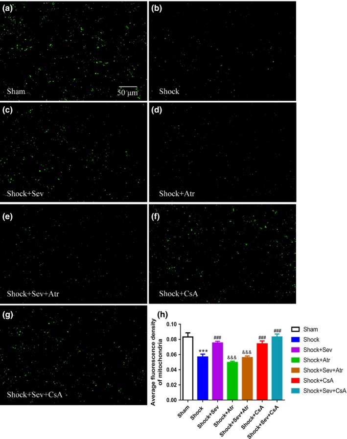
Methods: Sprague Dawley rats were subjected to hemorrhage shock for 60 min and then exposed to 2.4% sevoflurane for 30 min at the instant of reperfusion. Additionally, an opener (atractyloside) or an inhibitor (cyclosporine A) of mPTP was used in the animal model before sevoflurane postconditioning. Rats were randomly assigned into groups of Sham, Shock, Shock+Sevoflurane, Shock+Atractyloside, Shock+Sevoflurane+Atractyloside, Shock+Cyclosporin A, and Shock+Sevoflurane+Cyclosporin A treatment. Rat behavior was assessed for 5 days by Morris water maze 72 hr after surgery, and then hippocampus CA1 region was immunohistochemically stained. Brains were harvested 24 hr after surgery to detect the protein expression levels of Bcl-2, Bax, and cytochrome C by Western blot, the changes of mPTP opening, and mitochondrial membrane potential (MMP).
Results: We found that sevoflurane postconditioning significantly improved rats' spatial learning and memory ability, down-regulated the expression of Bax, cytochrome C, and caspase-3, up-regulated the expression of Bcl-2, decreased the mPTP opening, and increased the MMP. The neuroprotective effect of sevoflurane postconditioning was abolished by atractyloside, but cyclosporin A played the similar protective role as sevoflurane postconditioning.
Conclusion: These findings proved that sevoflurane postconditioning improved spatial learning and memory ability in hemorrhage shock and resuscitation rats, the mechanism of which may be related to block mPTP opening, increase MMP, and reduce neuron apoptosis in the hippocampus.
Keywords: apoptosis; hemorrhagic shock and resuscitation; mitochondrial membrane potential; mitochondrial permeability transition pore; sevoflurane postconditioning; spatial learning and memory.
© 2019 The Authors. Brain and Behavior published by Wiley Periodicals, Inc.
Conflict of interest statement
The authors declare no conflict of interests.
Figures


Similar articles
-
Sevoflurane postconditioning improves the spatial learning and memory impairments induced by hemorrhagic shock and resuscitation through suppressing IRE1α-caspase-12-mediated endoplasmic reticulum stress pathway.Neurosci Lett. 2018 Oct 15;685:160-166. doi: 10.1016/j.neulet.2018.08.035. Epub 2018 Aug 26. Neurosci Lett. 2018. PMID: 30157449
-
Postconditioning with sevoflurane ameliorates spatial learning and memory deficit after hemorrhage shock and resuscitation in rats.J Surg Res. 2016 Dec;206(2):307-315. doi: 10.1016/j.jss.2016.08.026. Epub 2016 Aug 11. J Surg Res. 2016. PMID: 27884324
-
Sevoflurane postconditioning improves long-term learning and memory of neonatal hypoxia-ischemia brain damage rats via the PI3K/Akt-mPTP pathway.Brain Res. 2016 Jan 1;1630:25-37. doi: 10.1016/j.brainres.2015.10.050. Epub 2015 Nov 2. Brain Res. 2016. PMID: 26541582
-
Recent progress in the use of mitochondrial membrane permeability transition pore in mitochondrial dysfunction-related disease therapies.Mol Cell Biochem. 2021 Jan;476(1):493-506. doi: 10.1007/s11010-020-03926-0. Epub 2020 Sep 30. Mol Cell Biochem. 2021. PMID: 33000352 Review.
-
The mitochondrial permeability transition pore in cell death: A promising drug binding bioarchitecture.Med Res Rev. 2020 Mar;40(2):811-817. doi: 10.1002/med.21635. Epub 2019 Oct 16. Med Res Rev. 2020. PMID: 31617227 Review.
Cited by
-
Preconditioning and Posttreatment Strategies in Neonatal Hypoxic-Ischemic Encephalopathy: Recent Advances and Clinical Challenges.Mol Neurobiol. 2025 Aug;62(8):10020-10044. doi: 10.1007/s12035-025-04896-4. Epub 2025 Apr 3. Mol Neurobiol. 2025. PMID: 40178781 Review.
-
THE MITOCHONDRIAL DIVISION INHIBITOR MDIVI-1 PROTECTED ORGAN FUNCTION AND EXTENDED THE TREATMENT WINDOW IN RATS WITH UNCONTROLLED HEMORRHAGIC SHOCK.Shock. 2025 Mar 1;63(3):474-486. doi: 10.1097/SHK.0000000000002459. Epub 2024 Aug 26. Shock. 2025. PMID: 39185710 Free PMC article.
-
Sevoflurane postconditioning reduces myocardial ischemia reperfusion injury-induced necroptosis by up-regulation of OGT-mediated O-GlcNAcylated RIPK3.Aging (Albany NY). 2020 Nov 20;12(24):25452-25468. doi: 10.18632/aging.104146. Epub 2020 Nov 20. Aging (Albany NY). 2020. PMID: 33231560 Free PMC article.
-
Sevoflurane Induces Neurotoxicity in the Animal Model with Alzheimer's Disease Neuropathology via Modulating Glutamate Transporter and Neuronal Apoptosis.Int J Mol Sci. 2022 Jun 2;23(11):6250. doi: 10.3390/ijms23116250. Int J Mol Sci. 2022. PMID: 35682930 Free PMC article.
-
Kapβ2 Reverses Sevoflurane-Induced Hydrogel Phase Transition of hnRNPA2/B1-SG in Hypoxic Primary Rat Hippocampal Neurons.CNS Neurosci Ther. 2025 Aug;31(8):e70532. doi: 10.1111/cns.70532. CNS Neurosci Ther. 2025. PMID: 40776395 Free PMC article.
References
-
- Akopova, O. V. (2008). The role of mitochondrial permeability transition pore in transmembrane Ca2+‐exchange in mitochondria. Ukrainskii Biokhimicheskii Zhurnal, 80(3), 40–47. - PubMed
-
- Basso, E. , Petronilli, V. , Forte, M. A. , & Bernardi, P. (2008). Phosphate is essential for inhibition of the mitochondrial permeability transition pore by cyclosporin A and by cyclophilin D ablation. Journal of Biological Chemistry, 283(39), 26307–26311. 10.1074/jbc.C800132200 - DOI - PMC - PubMed
-
- Bonnet, S. , Archer, S. L. , Allalunis‐Turner, J. , Haromy, A. , Beaulieu, C. , Thompson, R. , … Michelakis, E. D. (2007). A mitochondria‐K+ channel axis is suppressed in cancer and its normalization promotes apoptosis and inhibits cancer growth. Cancer Cell, 11(1), 37–51. 10.1016/j.ccr.2006.10.020 - DOI - PubMed
Publication types
MeSH terms
Substances
Grants and funding
LinkOut - more resources
Full Text Sources
Medical
Research Materials
