. 2019 Dec;47(8):1049-1071.
doi: 10.1177/0192623319879903.

Endocrine Disruption and Reproductive Pathology

Affiliations

Endocrine Disruption and Reproductive Pathology

Scott M Belcher et al. Toxicol Pathol. 2019 Dec.

Abstract

During the past 20 years, investigations involving endocrine active substances (EAS) and reproductive toxicity have dominated the landscape of ecotoxicological research. This has occurred in concert with heightened awareness in the scientific community, general public, and governmental entities of the potential consequences of chemical perturbation in humans and wildlife. The exponential growth of experimentation in this field is fueled by our expanding knowledge into the complex nature of endocrine systems and the intricacy of their interactions with xenobiotic agents. Complicating factors include the ever-increasing number of novel receptors and alternate mechanistic pathways that have come to light, effects of chemical mixtures in the environment versus those of single EAS laboratory exposures, the challenge of differentiating endocrine disruption from direct cytotoxicity, and the potential for transgenerational effects. Although initially concerned with EAS effects chiefly in the thyroid glands and reproductive organs, it is now recognized that anthropomorphic substances may also adversely affect the nervous and immune systems via hormonal mechanisms and play substantial roles in metabolic diseases, such as type 2 diabetes and obesity.

Keywords: endocrine disrupters; environmental toxicology; fish pathology; hormonal carcinogenesis; nonhuman primate; reproductive system; rodent pathology.


Conflict of interest statement

Declaration of Conflicting Interests

The author(s) declared no potential conflicts of interest with respect to the research, authorship, and/or publication of this article.

Figures

Figure 1.
Figure 1.
Adverse outcome pathway (AOP) network for chemicals that disrupt fetal development of the male reproductive tract. The predominant pathways include reduction in androgen receptor (AR)-dependent gene/protein expression; however, additional pathways including insulin-like hormone 3 (Insl3), estrogen receptor, and aryl hydrocarbon receptor (AhR) are known to adversely affect male development. DHT indicates dihydrotestosterone; HMG-CoA, 3-hydroxy-3-methyl-glutaryl coenzyme A; MIE, molecular initiating event.
Figure 2.
Figure 2.
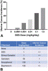
Diethylstilbestrol (DES) as a model for weaker environmental estrogens. Mice were treated on neonatal days 1 to 5 with increasing doses of DES (panel A) or environmental chemicals (panel B) and uterine adenocarcinoma incidence determined at 12 months of age.
Figure 3.
Figure 3.
Aberrant cell types in mouse and human uterine carcinoma. A and B, Uterine adenocarcinoma lesion from an 18-month-old mouse that was neonatally exposed to GEN. Abnormal cells uniformly express Six1 (panel A) and have luminal (CK18, brown, asterisk), basal (CK14, teal, arrowhead), or mixed (CK14/CK18 coexpression, forest green, arrows) features based on morphology and dual CK14/CK18 labeling (panel B). Images show serial sections. C and D, Human uterine carcinoma with Six1 expression (panel C) and cells coexpressing CK14 and CK18 embedded within the neoplastic lesion (panel D). Images shows cells within the same region, but are not serial sections. All slides were immunostained for Six1 or doubled immunostained for CK14 and CK18. Original objective ×40. CK indicates cytokeratin; GEN, genistein; Six 1, sine oculis homeobox transcription factor 1.
Figure 4.
Figure 4.
The CLARITY-BPA study design. The CLARITY-BPA study design is shown graphically with dosing initiated at GD6 through PND21 (stop-dose) or continuously until the indicated time of sacrifice (sac) and tissue collection. CLARITY-BPA indicates Consortium Linking Academic and Regulatory Insights on BPA Toxicity; GD, gestational day; LV, left ventricle; PND, postnatal day; Sac, sacrifice/necropsy.
Figure 5.
Figure 5.
Normal endometrial histology of the cynomolgus monkey. A and F, Atrophic endometrium, with sparse straight glands, cuboidal glandular epithelium, and compact stroma. B and G, Follicular-phase endometrium, with pseudostratified glandular epithelium, and straight to slightly coiled glands in an edematous stroma. C and H, Periovulatory endometrium, with distinct subnuclear vacuoles. D and I, Luteal-phase endometrium, with saw-toothed glands and prominent spiral arteries. E and J, Menstrual phase, with compact stroma, sloughing, hemorrhage, and apoptosis.
Figure 6.
Figure 6.
Histologic changes characteristic of estrogens, progestogens, and selective estrogens in the endometrium of cynomolgus monkeys. A and E, Ovariectomy-induced endometrial atrophy. B and F, Estrogen-induced irregular endometrial glandular hyperplasia. C and G, Estrogen- + progestogen-induced glandular atrophy, stromal hyperplasia, and infiltration by endometrial granular leukocytes. D and H, Stromal fibrosis and cystic change induced by a selective estrogen (tamoxifen).
Figure 7.
Figure 7.
The epithelial plaque response in the endometrium of a cynomolgus monkey (reprinted with permission from Cline et al). This lesion occurs in the luteal phase of thecycle and is an incidental finding. A, a plaque of endometrial surfaceepithelium covering most of the luminal surface, with “saw-toothed”luteal-phase endometrial glands in the deeper endometrium. B, highermagnification of epithelial plaque cells, showing large round to polygonalcells with clear cytoplasm surrounding a blood vessel. C, immunohistochemicalstaining for cleaved caspase 3, indicating apoptosis.
Figure 8.
Figure 8.
Squamous metaplasia of the endocervix in a cynomolgus monkey. This benign lesion is induced by physiologic or pharmacologic estrogens. Arrows indicate nests of squamous cells beneath the normal pseudostratified columnar ciliated epithelium of the endocervix.
Figure 9.
Figure 9.
Papillomavirus-induced lesions of the vagina and cervix in cynomolgus monkeys. A, Normal, (B) epithelial atypia, (C) basal hyperplasia, and (D) atypia bordering on squamous cell carcinoma.
Figure 10.
Figure 10.
Condylomatous lesion of the genital mucosa in a cynomolgus monkey, consisting of regular hyperplasia without atypia, and not associated with papillomavirus infection.
Figure 11.
Figure 11.
Seasonal testicular atrophy in rhesus macaques. A, Normal testis, winter. B, Testicular atrophy, summer. C, Normal epididymis, winter. D, Hypospermia, summer.
Figure 12.
Figure 12.
Prostatic basal cell hyperplasia in a cynomolgus monkey. Hyperplastic glands are on the lower leftside of the image and consist of basophilic, multilayered to solid, small,polygonal cells with scant cytoplasm filling the glands, in contrast to thesingle-layered, simple columnar epithelium of adjacent normal glands.
Figure 13.
Figure 13.
Study site map, state of North Carolina. Collection sites were selected as follows: 8 point sources (red), defined by direct discharge into water; 9 nonpoint sources (yellow), with indirect diffusion into water; and 3 reference sites (blue), with no apparent discharge upstream. Up to 10 male black bass (Micropterus spp), 10 male sunfish (Lepomis spp), and 10 male catfish (Ictaluridae) were collected at each site whenever possible.
Figure 14.
Figure 14.
Representative cross-sections of testis from each fish were scored for intersex severity, using the method developed by Blazer et al. Oocytes observed within the seminiferous tubules were predominantly in the previtellogenic, chromatin nucleolus stage (arrows). A, Severity rank 1, single oocyte observed at × 10 objective magnification. B, Severity rank 2, more than 1 oocyte per field of view, but oocytes not closely associated. C, Severity rank 3, 2 to 5 closely associated oocytes. D, Severity rank 4, clusters of more than 5 closely associated oocytes. Hematoxylin and eosin staining.

References

    1. Mai CT, Isenburg J, Langlois PH, et al. Population-based birth defects data in the united states, 2008 to 2012: presentation of state-specific data and descriptive brief on variability of prevalence. Birth Defects Res A Clin Mol Teratol. 2015;103(11):972–993. - PMC - PubMed
    1. Paulozzi LJ. International trends in rates of hypospadias and cryptorchidism. Environ Health Perspect. 1999;107(4):297–302. - PMC - PubMed
    1. Macleod DJ, Sharpe RM, Welsh M, et al. Androgen action in the masculinization programming window and development of male reproductive organs. Int J Androl. 2010;33(2):279–287. - PubMed
    1. Wilson VS, Blystone CR, Hotchkiss AK, Rider CV, Gray LE JR. Diverse mechanisms of anti-androgen action: impact on male rat reproductive tract development. Int J Androl. 2008;31(2):178–187. - PubMed
    1. Blystone CR, Lambright CS, Howdeshell KL, et al. Sensitivity of fetal rat testicular steroidogenesis to maternal prochloraz exposure and the underlying mechanism of inhibition. Toxicol Sci. 2007; 97(2):512–519. - PubMed

Publication types

MeSH terms

LinkOut - more resources