. 2020 Feb;165(2):345-354.
doi: 10.1007/s00705-019-04470-z. Epub 2019 Dec 13.

miR-142a-3p promotes the proliferation of porcine hemagglutinating encephalomyelitis virus by targeting Rab3a

Affiliations

miR-142a-3p promotes the proliferation of porcine hemagglutinating encephalomyelitis virus by targeting Rab3a

Peng Fan et al. Arch Virol. 2020 Feb.

Abstract

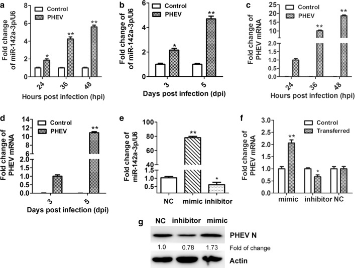
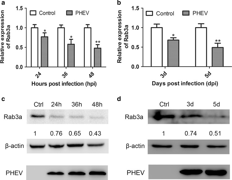
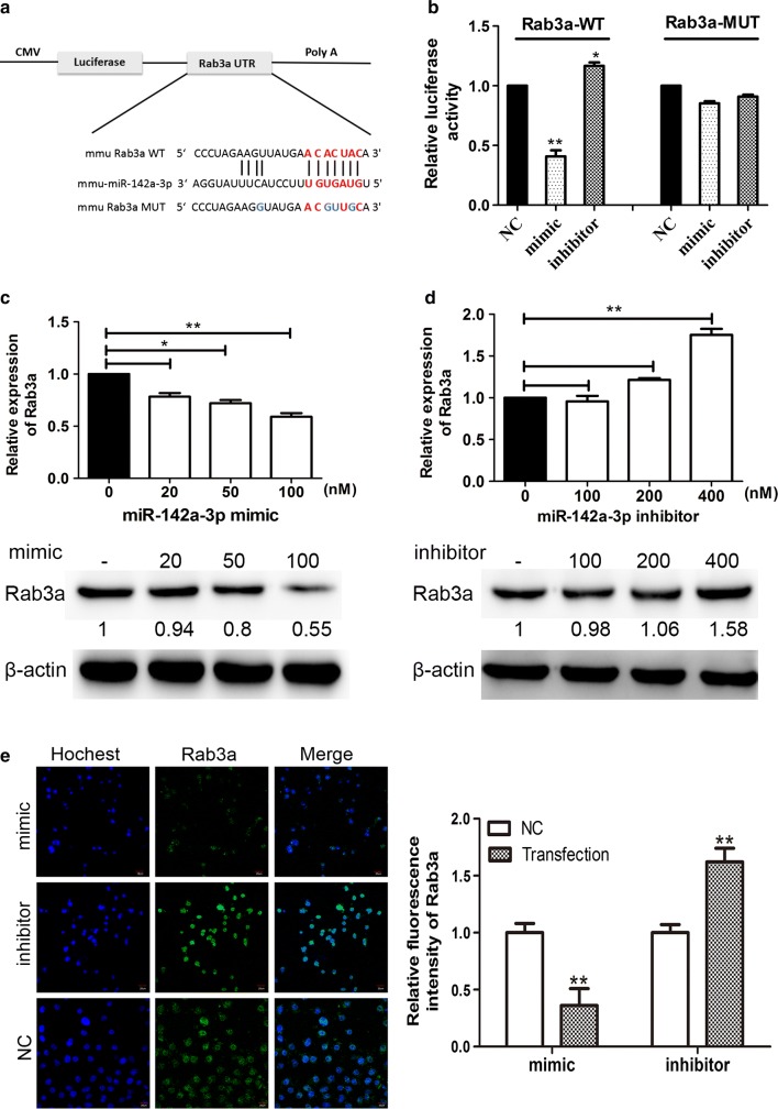
Porcine hemagglutinating encephalomyelitis virus (PHEV) is a typical neurotropic coronavirus that mainly invades the central nervous system (CNS) in piglets and causes vomiting and wasting disease. Emerging evidence suggests that PHEV alters microRNA (miRNA) expression profiles, and miRNA has also been postulated to be involved in its pathogenesis, but the mechanisms underlying this process have not been fully explored. In this study, we found that PHEV infection upregulates miR-142a-3p RNA expression in N2a cells and in the CNS of mice. Downregulation of miR-142a-3p by an miRNA inhibitor led to a significant repression of viral proliferation, implying that it acts as a positive regulator of PHEV proliferation. Using a dual-luciferase reporter assay, miR-142a-3p was found to bind directly bound to the 3' untranslated region (3'UTR) of Rab3a mRNA and downregulate its expression. Knockdown of Rab3a expression by transfection with an miR-142a-3p mimic or Rab3a siRNA significantly increased PHEV replication in N2a cells. Conversely, the use of an miR-142a-3p inhibitor or overexpression of Rab3a resulted in a marked restriction of viral production at both the mRNA and protein level. Our data demonstrate that miR-142a-3p promotes PHEV proliferation by directly targeting Rab3a mRNA, and this provides new insights into the mechanisms of PHEV-related pathogenesis and virus-host interactions.


Conflict of interest statement

The authors declare that they have no conflict of interest.

Figures

Fig. 1
Fig. 1
miR-142a-3p is upregulated during PHEV infection in vitro and in vivo. (a) PHEV-infected N2a cells were collected at different time points. The expression of miR-142a-3p was measured by qRT-PCR. (b) miR-142a-3p expression in mouse brain tissues after infection with PHEV was measured by qRT-PCR. (c) Cells that were pre-treated as indicated above were analyzed by qRT-PCR to measure the level of PHEV mRNA. (d) PHEV mRNA expression in mouse brain tissues after infection with PHEV was measured by qRT-PCR. (e) N2a cells were transfected with miR-142a-3p mimic (100 nM), inhibitor (400 nM), or NC (100 nM). Expression of miR-142a-3p was then measured by qRT-PCR, with the housekeeping gene U6 used as an internal control for normalization. (f) N2a cells were transfected with the miR-142a-3p mimic, inhibitor, or NC for 12 h, followed by PHEV infection for 24 h. Lysates were harvested and tested by qRT-PCR. (g) Cells were pre-treated as indicated above, lysed, and analyzed using a Western blotting assay to determine the level of PHEV N protein. Data were normalized to β-actin. All of the data are representative of at least three independent experiments (*, p < 0.05; **, p < 0.01)
Fig. 2
Fig. 2
Rab3a expression is inhibited during PHEV infection in vitro and in vivo. (a) N2a cells were mock infected (control) or infected with PHEV for the indicated times. Endogenous Rab3a mRNA expression was measured by qRT-PCR and compared to that of the control. (b) Mice were mock infected (control) or infected with PHEV. At 3 or 5 dpi, the cerebral cortexes were harvested and Rab3a mRNA was quantitated by qRT-PCR and normalized to GAPDH. (c) Lysates of mock-infected N2a cells or cells that were infected with PHEV were analyzed by Western blotting using primary antibodies against Rab3a or β-actin. The data were normalized to β-actin. (d) Lysates from cerebral cortexes of mice that were pre-treated as indicated above were analyzed by Western blotting and normalized to β-actin. All of the data are representative of at least three independent experiments (*, p < 0.05; **, p < 0.01)
Fig. 3
Fig. 3
Rab3a is a target gene of miR-142a-3p. (a) Schematic representation of WT or MUT luciferase reporter of Rab3a mRNA 3′UTR. The frame and letters in blue indicate the point mutant. (b) HEK293T cells were cotransfected with Rab3a-WT or Rab3a-MUT reporter constructs and the indicated oligonucleotides (100 nM mimic, 400 nM inhibitor, or 100 nM NC) for 24 h. The cells were harvested, and Renilla and firefly luciferase activity was measured. (c) The mRNA and protein levels of Rab3a in N2a cells transfected with miR-142a-3p mimic were determined by RT-PCR and Western blotting. (d) The expression of Rab3a was measured in N2a cells after transfection with miR-142a-3p inhibitor. (e) Representative micrograph of immunofluorescence analysis for Rab3a expression (green) in N2a cells transfected with the miR-142a-3p mimic- or miR-142a-3p inhibitor. The cell nucleus was stained with Hochest33342 (blue). Quantitative analysis of the Rab3a staining is shown on the right. The bars indicate 20 μm. All of the data are representative of at least three independent experiments (*, p < 0.05; **, p < 0.01)
Fig. 4
Fig. 4
Rab3a plays a passive regulatory role in PHEV proliferation. (a) The expression of Rab3a mRNA in N2a cells transfected with 50 nM siRab3a or siNC was measured by qRT-PCR. Normal cells were transfected without any siRNA. (b) The protein levels of Rab3a in N2a cells transfected with siRNA were determined by Western blotting. (c) The levels of viral genomic RNA were measured after transfecting with Rab3a siRNA. Untransfected cells were used as a control. (d) Western blotting was performed to examine the expression of PHEV N protein. (e and f) The expression of Rab3a in cells that transfected with Rab3a-GFP was examined by qRT-PCR (e) and Western blotting (f). (g and h) The viral genomic RNA and PHEV N protein levels in Rab3a-GFP-transfected cells were determined. All of the data are representative of at least three independent experiments (*, p < 0.05; **, p < 0.01; ***, p <0.001)

Similar articles

Cited by

References

    1. Dong B, Lu H, Zhao K, Liu W, Gao W, Lan Y, Zhao J, Tang B, Song D, He W, Gao F. Identification and genetic characterization of porcine hemagglutinating encephalomyelitis virus from domestic piglets in China. Arch Virol. 2014;159:2329–2337. doi: 10.1007/s00705-014-2070-y. - DOI - PMC - PubMed
    1. Gao W, Zhao K, Zhao C, Du C, Ren W, Song D, Lu H, Chen K, Li Z, Lan Y, Xie S, He W, Gao F. Vomiting and wasting disease associated with hemagglutinating encephalomyelitis viruses infection in piglets in Jilin, China. Virol J. 2011;8:130. doi: 10.1186/1743-422X-8-130. - DOI - PMC - PubMed
    1. Roe CK, Alexander TJ. A disease of nursing pigs previously unreported in Ontario. Can J Comp Med Vet Sci. 1958;22:305–307. - PMC - PubMed
    1. Greig AS, Mitchell D, Corner AH, Bannister GL, Meads EB, Julian RJ. A Hemagglutinating Virus Producing Encephalomyelitis in Baby Pigs. Can J Comp Med Vet Sci. 1962;26:49–56. - PMC - PubMed
    1. Lan Y, Zhao K, Wang G, Dong B, Zhao J, Tang B, Lu H, Gao W, Chang L, Jin Z, Gao F, He W. Porcine hemagglutinating encephalomyelitis virus induces apoptosis in a porcine kidney cell line via caspase-dependent pathways. Virus Res. 2013;176:292–297. doi: 10.1016/j.virusres.2013.05.019. - DOI - PMC - PubMed

LinkOut - more resources