Review
. 2021 Sep;24(3):331-336.
doi: 10.1007/s40477-019-00418-w. Epub 2019 Dec 13.

Thoracic outlet syndrome: a rare case with bilateral cervical ribs and bilateral anterior scalene hypertrophy

Affiliations
Review

Thoracic outlet syndrome: a rare case with bilateral cervical ribs and bilateral anterior scalene hypertrophy

Renato Farina et al. J Ultrasound. 2021 Sep.

Abstract

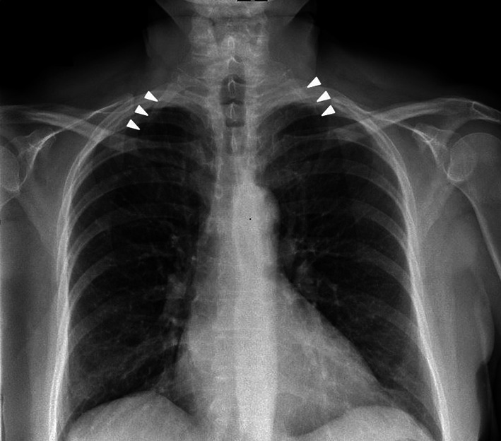
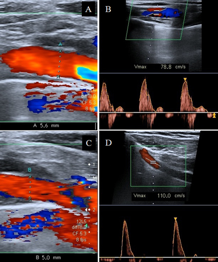
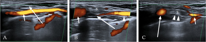
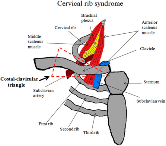
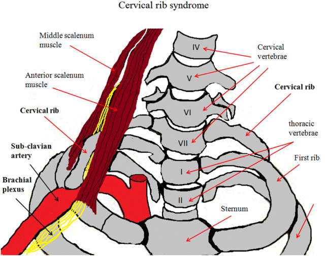
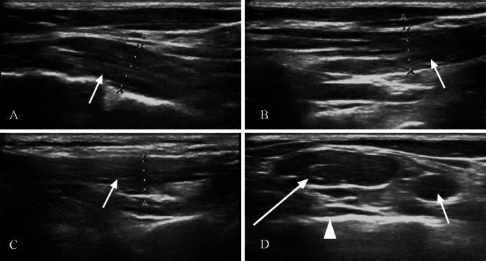
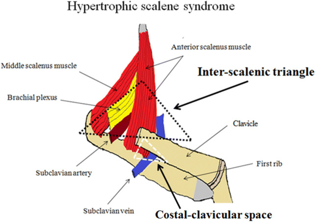
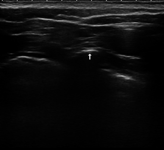
Thoracic outlet syndrome (TOS) is a rare neurovascular disorder generally caused by the presence of a cervical rib or hypertrophic scalene anterior muscle that can compress the brachial plexus and/or subclavian vessels. In the vascular form, the symptoms are caused by the compression of the artery and/or the subclavian vein. In the first case, the compression is caused by the cervical rib and leads to hypo-perfusion with cooling and cyanosis of the upper limb, while in the second case, the compression is caused by the anterior scalene muscle and leads to congestion, cyanosis, swelling and pain in the higher limb. In this paper, we describe a case with the simultaneous presence of a bilateral cervical rib and bilateral hypertrophy of the anterior scalene muscle. TOS diagnosis is based on neurological, clinical and instrumental tests, such as chest radiography and color Doppler ultrasonography. The treatment of these patients can be surgical or conservative.

Keywords: Anterior scalene; Color Doppler; Thoracic outlet syndrome.


Conflict of interest statement

The authors declare that they have no conflict of interest.

Figures

Fig. 1
Fig. 1
Standard chest X-ray. In this image, the right cervical rib (short arrow) and the left cervical rib (arrow heads) are easily identified
Fig. 2
Fig. 2
Sub-clavicular ultrasound scan. The color Doppler and Duplex ultrasonography study, performed in the middle section of the right subclavian artery with lowered arms, shows: a a regular diameter (5.6 mm) (arrow) and b regular systolic peak velocity (78.8 cm/s). The ultrasound scan, performed with arms raised, shows: c a slight diameter reduction (5 mm) (arrow) and d a slight increase in the systolic velocity (110 cm/s)
Fig. 3
Fig. 3
Power Doppler of the inter-scalene triangle. a The ultrasound scan, performed with lowered arms, shows a regular course and morphology of the right subclavian vein (short arrow). The anterior scalene muscle (long arrow). b The ultrasound scan, performed with arms at 90°, shows significant stenosis of the right subclavian vein (long arrow) and congestion in the pre-stenotic tract (short arrow) caused by the anterior scalene muscle (S). The stenotic tract of the subclavian vein (arrow heads). c In the ultrasound scan performed with arms raised, the anterior scalene muscle (arrow heads) can be seen causing the complete stenosis of the right subclavian vein, with dilatation of the pre-stenotic tract (long arrow) and diameter reduction in the post-stenotic tract (short arrow)
Fig. 4
Fig. 4
The costal-clavicular triangle, the space involved in cervical rib syndrome, consists anteriorly of the middle third of the clavicle, posteromedially from the first rib and posterolaterally from the upper profile of the scapula
Fig. 5
Fig. 5
In cervical rib syndrome, the cervical rib may cause compression of the subclavian artery and brachial plexus
Fig. 6
Fig. 6
B-Mode ultrasound. The laterocervical, longitudinal scans show: a anterior scalene muscle (arrow). b Medium scalene muscle (arrow). c Posterior scalene muscle. d In this scan, the inter-scalene triangle is highlighted, along with the anterior scalene muscle (long arrow), the subclavian vein (short arrow) and the first rib (arrow head)
Fig. 7
Fig. 7
The inter-scalene triangle, the anatomical space involved in the hypertrophic scalene syndrome, is formed anteriorly by the anterior scalene muscle, posteriorly by the medial scalene muscle and inferiorly from the medial face of the first rib (black triangle). In the costal-clavicular space (white triangle), compression of the subclavian vein generally occurs following hypertrophy of the anterior scalene muscle
Fig. 8
Fig. 8
B-mode ultrasound: transverse subclavian scan showing a cervical rib (arrow)

References

    1. Klaassen Z, Sorenson E, Tubbs RS, et al. Thoracic outlet syndrome: a neurological and vascular disorder. Clin Anat. 2014;27(5):724–732. doi: 10.1002/ca.22271. - DOI - PubMed
    1. Jones MR, Prabhakar A, Viswanath O, et al. Thoracic outlet syndrome: a comprehensive review of pathophysiology, diagnosis, and treatment. Pain Ther. 2019;8(1):5–18. doi: 10.1007/s40122-019-0124-2. - DOI - PMC - PubMed
    1. Jiang S, Shen H, Tan WQ, et al. Arterial thoracic outlet syndrome caused by cervical ribs-an unusual case report. Medicine (Baltimore) 2019;98(11):e14778. doi: 10.1097/MD.0000000000014778. - DOI - PMC - PubMed
    1. Chang KZ, Likes K, Davis K, et al. The significance of cervical ribs in thoracic outlet syndrome. J Vasc Surg. 2013;57(3):771–775. doi: 10.1016/j.jvs.2012.08.110. - DOI - PubMed
    1. Ozoa G, Alves D, Fish DE. Thoracic outlet syndrome. Phys Med Rehabil Clin N Am. 2011;22(3):473–483. doi: 10.1016/j.pmr.2011.02.010. - DOI - PubMed

MeSH terms