. 2019 Dec 13;15(12):e1008526.
doi: 10.1371/journal.pgen.1008526. eCollection 2019 Dec.

Genome wide analysis reveals heparan sulfate epimerase modulates TDP-43 proteinopathy

Affiliations

Genome wide analysis reveals heparan sulfate epimerase modulates TDP-43 proteinopathy

Nicole F Liachko et al. PLoS Genet. .

Abstract

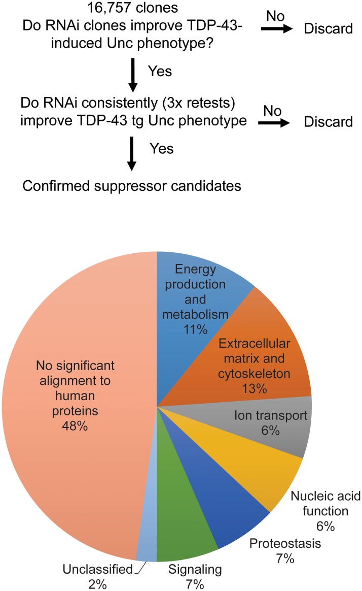
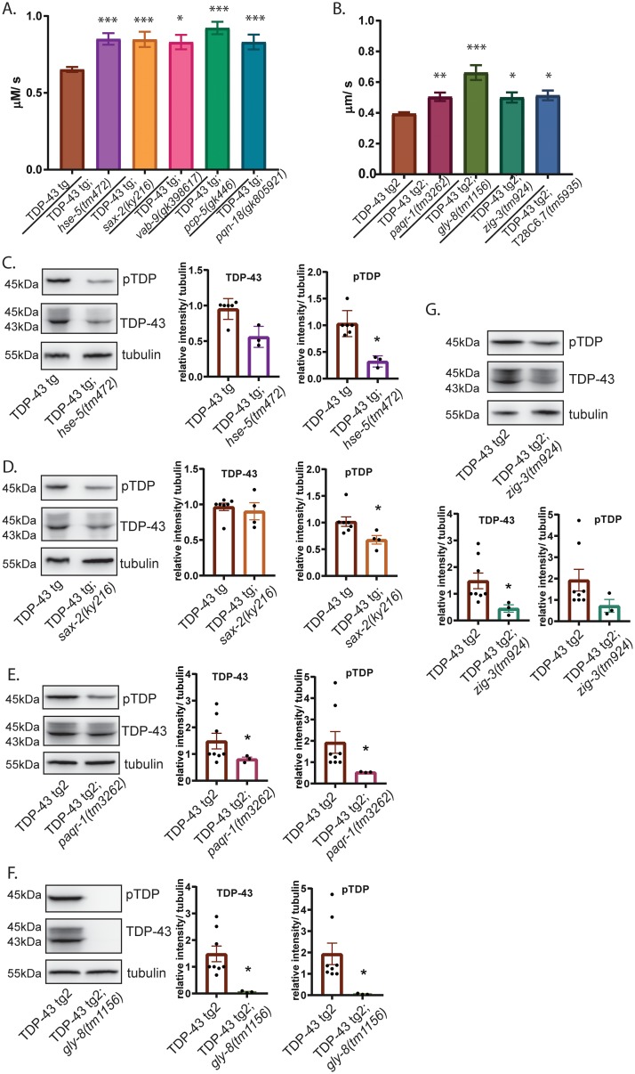
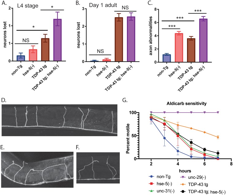
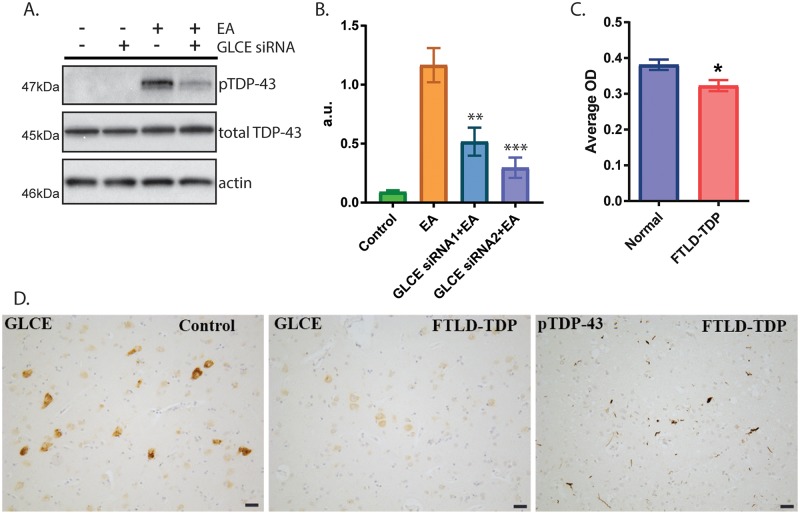
Pathological phosphorylated TDP-43 protein (pTDP) deposition drives neurodegeneration in amyotrophic lateral sclerosis (ALS) and frontotemporal lobar degeneration (FTLD-TDP). However, the cellular and genetic mechanisms at work in pathological TDP-43 toxicity are not fully elucidated. To identify genetic modifiers of TDP-43 neurotoxicity, we utilized a Caenorhabditis elegans model of TDP-43 proteinopathy expressing human mutant TDP-43 pan-neuronally (TDP-43 tg). In TDP-43 tg C. elegans, we conducted a genome-wide RNAi screen covering 16,767 C. elegans genes for loss of function genetic suppressors of TDP-43-driven motor dysfunction. We identified 46 candidate genes that when knocked down partially ameliorate TDP-43 related phenotypes; 24 of these candidate genes have conserved homologs in the human genome. To rigorously validate the RNAi findings, we crossed the TDP-43 transgene into the background of homozygous strong genetic loss of function mutations. We have confirmed 9 of the 24 candidate genes significantly modulate TDP-43 transgenic phenotypes. Among the validated genes we focused on, one of the most consistent genetic modifier genes protecting against pTDP accumulation and motor deficits was the heparan sulfate-modifying enzyme hse-5, the C. elegans homolog of glucuronic acid epimerase (GLCE). We found that knockdown of human GLCE in cultured human cells protects against oxidative stress induced pTDP accumulation. Furthermore, expression of glucuronic acid epimerase is significantly decreased in the brains of FTLD-TDP cases relative to normal controls, demonstrating the potential disease relevance of the candidate genes identified. Taken together these findings nominate glucuronic acid epimerase as a novel candidate therapeutic target for TDP-43 proteinopathies including ALS and FTLD-TDP.


Conflict of interest statement

The authors have declared that no competing interests exist.

Figures

Fig 1
Fig 1. Genome-wide RNAi screen identifies TDP-43 suppressors in several functional categories.
(A) Flowchart of the RNAi screen for modifiers of TDP-43-induced uncoordinated locomotion (Unc). (B) Percentage of suppressors identified in each functional group. Genes with no significant alignment within the human genome were not classified into functional groups.
Fig 2
Fig 2. Suppressors of TDP-43 improve C. elegans motor function and reduce levels of toxic TDP-43.
(A) Motor function of animals doubly homozygous for the human mutant TDP-43 (M337V) transgene, TDP-43 tg, and loss-of-function mutations for the indicated suppressor genes was assessed. Dispersal velocity of developmentally staged L4 larvae was measured by calculating the radial distance traveled from a designated central starting point over time, N>200 for all strains tested. Significance was evaluated using one-way analysis of variance with Tukey’s multiple comparison test among strains tested. ***p<0.0001, *p<0.01 versus TDP-43 tg. non-Tg (N2) animals move 3.712 μm/ sec. (B) To assay the effects of loss-of-function mutations in suppressor genes present on the same chromosome as the TDP-43 tg transgene (Chr IV), a second human mutant TDP-43 (A315T) transgene was utilized on a different chromosome (Chr II), TDP-43 tg2. Animals doubly homozygous for the TDP-43 tg2 transgene and loss-of-function mutations for the TDP-43 suppressor genes indicated were assessed for motor function as above, N>200 for all strains tested. ***p<0.0001, **p<0.001, *p<0.01 versus TDP-43 tg2. (C-G) Measurement of levels of total and phosphorylated TDP-43 protein by immunoblot. Data shown are representative of triplicate independent experiments. Graphs plot relative total TDP-43 or phosphorylated TDP-43 signal normalized to tubulin protein levels from three or more independent replicate experiments. Significance was evaluated using Mann-Whitney test between strains tested. *p<0.05.
Fig 3
Fig 3. hse-5 loss of function restores synaptic transmission in TDP-43 tg C. elegans.
(A) To assess neurodegeneration, GFP-labeled D-type GABAergic motor neurons were counted at L4 stage in vivo in living worms. TDP-43 tg; hse-5(tm472) animals lose slightly more neurons than TDP-43 tg alone, N>33 for all strains tested. Significance was evaluated using one-way analysis of variance with Tukey’s multiple comparison test among strains tested. *p<0.05, non-Tg versus TDP-43 tg and TDP-43 tg versus TDP-43 tg; hse-5(tm472), NS = not significant. (B) At day 1 adult, there are no differences in the number of D-type GABAergic motor neurons between TDP-43 tg versus TDP-43 tg; hse-5(tm472), N>33 for all strains tested. NS = not significant. p<0.0001 for non-Tg versus TDP-43tg and TDP-43 tg; hse-5(tm472). (C) hse-5(tm472) exhibit numerous axonal abnormalities including aberrant branching, looping, and inter-axonal connections. These non-canonical processes are increased in TDP-43 tg; hse-5(tm472) animals, N>33 for all strains tested. ***p<0.0001 for non-Tg versus hse-5(tm472), TDP-43tg, and TDP-43 tg; hse-5(tm472), and for TDP-43tg versus TDP-43 tg; hse-5(tm472). (D) Wild-type control axons ascend linearly from the ventral nerve cord without branching, looping, or inter-axonal connections. (E-F) TDP-43 tg; hse-5(tm472) axons frequently exhibit unusual branches, loops, or inter-axonal connections. (G) Pre-synaptic transmission was assessed using an aldicarb sensitivity assay. Animals were scored at the indicated time points for paralysis. Triplicate independent experiments are plotted. TDP-43 tg animals (orange diamond, p<0.005 at hours 3–7) are strongly resistant to aldicarb relative to non-Tg animals (blue circle) indicating decreased or defective synaptic transmission, while hse-5(tm472) animals (red square, p<0.05 at hours 3–4) are more modestly resistant to aldicarb. TDP-43 tg; hse-5(tm472) animals (black circle, p<0.05 at hours 3–5) have synaptic transmission restored to hse-5(tm472) levels. unc-29(e1072) (purple inverted triangle, p<0.05 at hours 3–7) and unc-31(e928) (green triangle, p<0.05 at hours 3 and 4) are controls for decreased aldicarb sensitivity. Significance was evaluated using two-way ANOVA with Tukey’s multiple comparisons test; p-values compared to non-Tg.
Fig 4
Fig 4. GLCE changes in mammalian cultured cells and in FTLD-TDP.
(A-B) Cultured HEK293 cells treated with ethacrynic acid (EA) accumulate robust levels of phosphorylated TDP-43. (A) Representative immunoblots of cells treated with GLCE-targeted siRNA exhibit reduced pTDP accumulation in the presence of EA. (B) Triplicate independent experiments were quantified and graphed. Significance was evaluated using one-way analysis of variance with Tukey’s multiple comparison test among strains tested. **p = 0.0029, ***p = 0.0002 versus EA treated. (C-D) GLCE immunoreactivity is reduced in patients with FTLD-TDP compared with normal controls. (C) Quantitation of GLCE immunoreactivity in the deep layers of the frontal cortex (mid-frontal gyrus) from 14 patients with FTLD-TDP and 12 normal controls. Significance was evaluated using an unpaired t-test, *p = 0.0116. (D) Representative images of GLCE immunostaining in the frontal cortex of patients and controls. pTDP-43 accumulation is apparent in FTLD-TDP patients in this same brain region. Scale bar = 50um.

References

    1. Tan RH, Ke YD, Ittner LM, Halliday GM. ALS/FTLD: experimental models and reality. Acta Neuropathologica. 2017:1–20. 10.1007/s00401-016-1666-6 - DOI - PubMed
    1. Mann DMA, Snowden JS. Frontotemporal lobar degeneration: Pathogenesis, pathology and pathways to phenotype. Brain Pathol. 2017. 10.1111/bpa.12486 . - DOI - PMC - PubMed
    1. Chornenkyy Y, Fardo DW, Nelson PT. Tau and TDP-43 proteinopathies: kindred pathologic cascades and genetic pleiotropy. Lab Invest. 2019. 10.1038/s41374-019-0196-y . - DOI - PMC - PubMed
    1. Wilson RS, Yu L, Trojanowski JQ, Chen EY, Boyle PA, Bennett DA, et al. TDP-43 pathology, cognitive decline, and dementia in old age. JAMA Neurol. 2013;70(11):1418–24. 10.1001/jamaneurol.2013.3961 - DOI - PMC - PubMed
    1. James BD, Wilson RS, Boyle PA, Trojanowski JQ, Bennett DA, Schneider JA. TDP-43 stage, mixed pathologies, and clinical Alzheimer’s-type dementia. Brain. 2016;139(11):2983–93. 10.1093/brain/aww224 - DOI - PMC - PubMed

Publication types

MeSH terms