. 2019 Dec 12;179(7):1566-1581.e16.
doi: 10.1016/j.cell.2019.11.022.

A Translation-Activating Function of MIWI/piRNA during Mouse Spermiogenesis

Affiliations

A Translation-Activating Function of MIWI/piRNA during Mouse Spermiogenesis

Peng Dai et al. Cell. .

Abstract

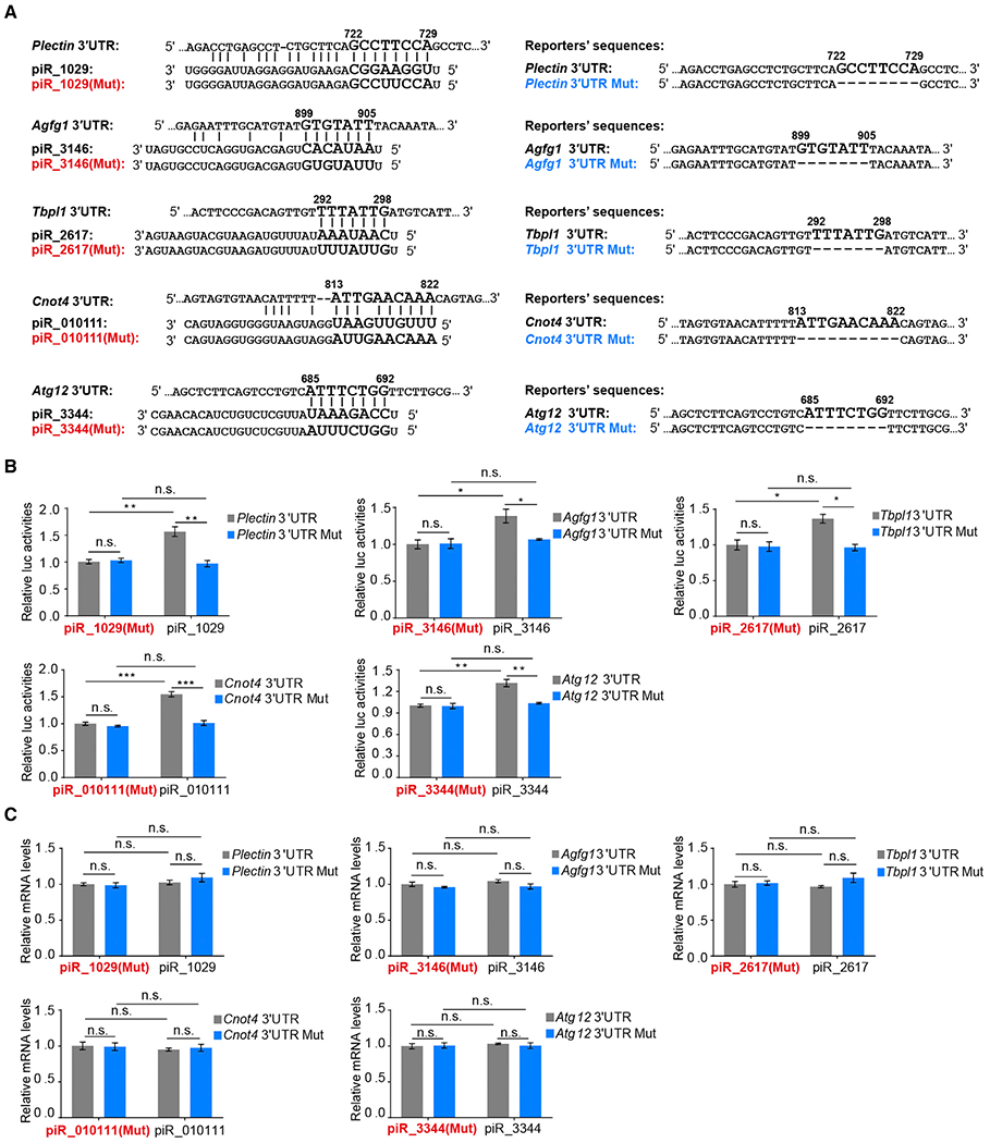
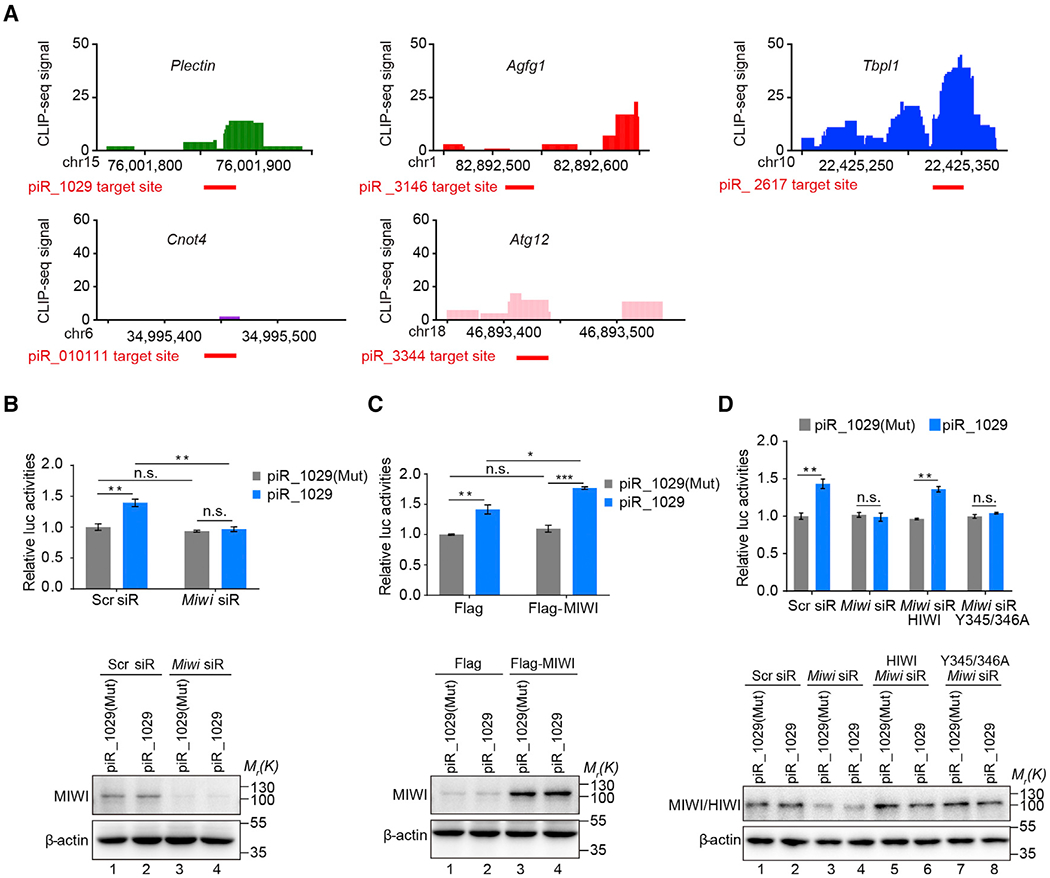
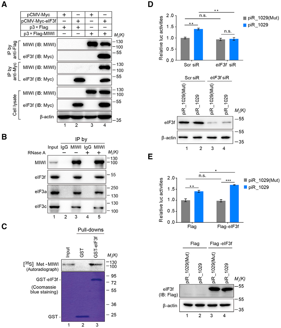
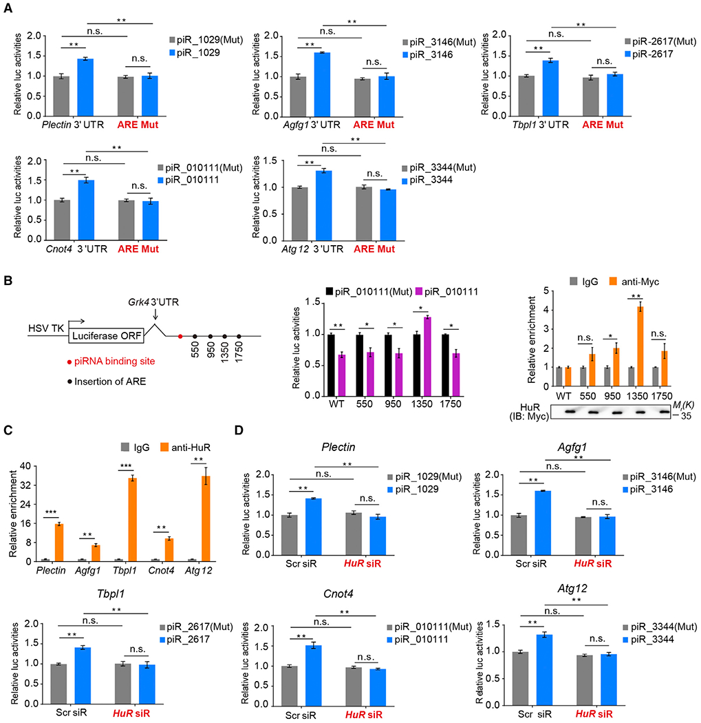
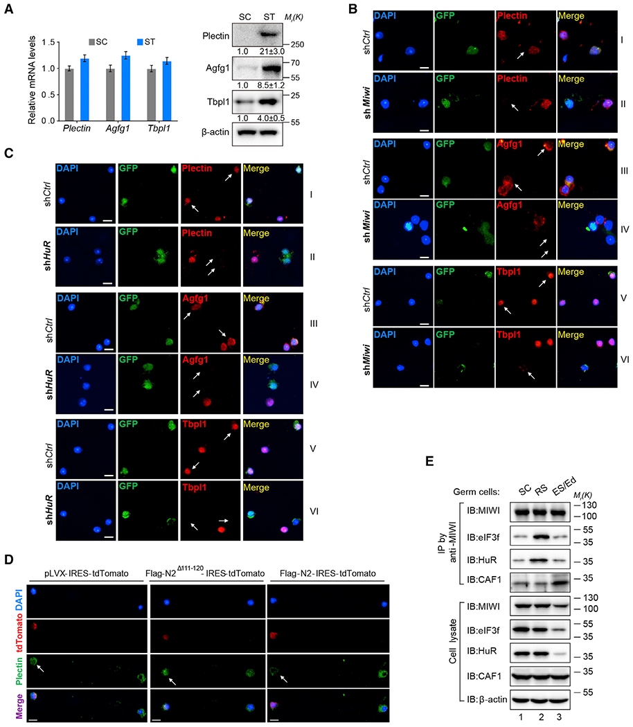
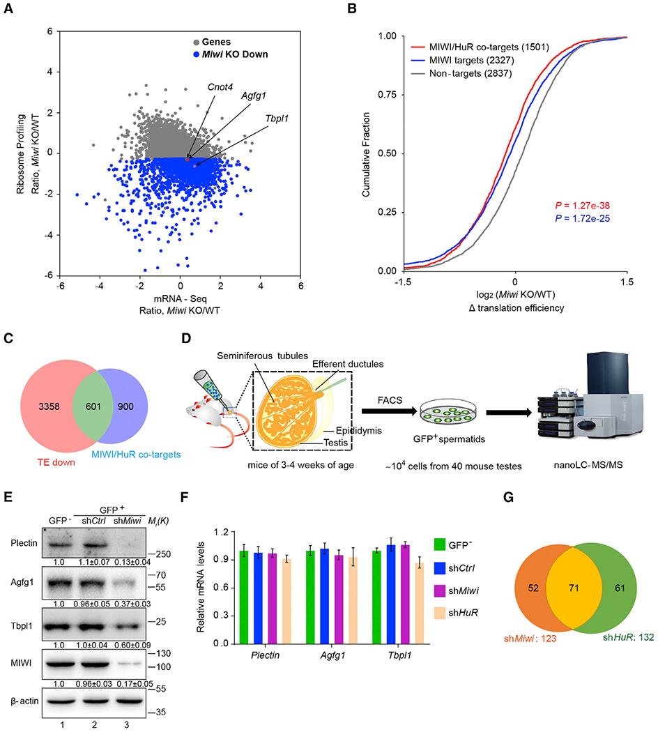
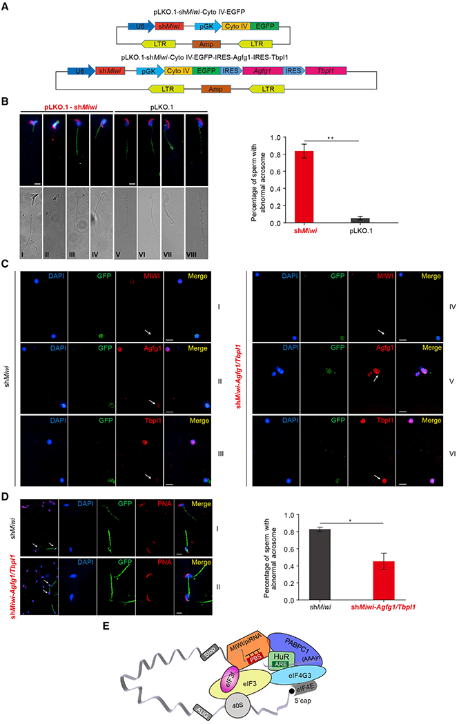
Spermiogenesis is a highly orchestrated developmental process during which chromatin condensation decouples transcription from translation. Spermiogenic mRNAs are transcribed earlier and stored in a translationally inert state until needed for translation; however, it remains largely unclear how such repressed mRNAs become activated during spermiogenesis. We previously reported that the MIWI/piRNA machinery is responsible for mRNA elimination during late spermiogenesis in preparation for spermatozoa production. Here we unexpectedly discover that the same machinery is also responsible for activating translation of a subset of spermiogenic mRNAs to coordinate with morphological transformation into spermatozoa. Such action requires specific base-pairing interactions of piRNAs with target mRNAs in their 3' UTRs, which activates translation through coupling with cis-acting AU-rich elements to nucleate the formation of a MIWI/piRNA/eIF3f/HuR super-complex in a developmental stage-specific manner. These findings reveal a critical role of the piRNA system in translation activation, which we show is functionally required for spermatid development.


Conflict of interest statement

DECLARATION OF INTERESTS

The authors declare no competing interests.

Figures

Figure 1.
Figure 1.. piRNAs Activate the Translation of Target mRNAs via Imperfectly Base Pairing with the 3′ UTRs
(A) Predicted piRNA regulatory elements at the 3′ UTRs of Plectin, Agfg1, Tbpl1, Cnot4, or Atg12 and the synthetic piRNAs, mutated piRNAs, and wild-type and mutated 3′ UTR Renilla luciferase reporters. (B) Dual-luciferase reporter assay showing that piRNAs promoted the activities of 3′ UTR reporters in GC-2spd (ts) cells, with mutated piRNAs serving as negative controls. (C) qPCR assay showing that piRNAs barely altered the levels of 3′ UTR reporter mRNAs. The average values ± SD of three separate experiments were plotted. *p < 0.05, **p < 0.01, ***p < 0.001, n.s., not significant. See also Figure S1.
Figure 2.
Figure 2.. MIWI Is Essential for piRNA-Induced Target Activation
(A) Verification of piRNA target sites by MIWI crosslinking immunoprecipitation (CLIP) in mouse spermatids. Top: the cluster of MIWI CLIP tags on the five validated piRNA-activated genes, with the scales in genes numbered following the dimension of the chromosomes; bottom: the red thick lines indicate piRNA target sites. (B and C) The effect of Miwi knockdown (B) or overexpression (C) on piR_1029-induced Plectin reporter activation in GC-2spd (ts) cells. Top: dual-luciferase reporter assay; bottom: western blotting assay of MIWI. (D) Ectopic Flag-HIWI, but not its piRNA-loading-deficient Y345/346A mutant, rescued piR_1029-induced Plectin reporter activation in Miwi siR-treated GC-2spd(ts) cells. Top: dual-luciferase reporter assay; bottom: western blotting assay of MIWI or HIWI. The average values ± SD of three separate experiments were plotted. *p < 0.05, **p < 0.01, ***p < 0.001, n.s., not significant. See also Figures S2A–S2D.
Figure 3.
Figure 3.. eIF3f, a MIWI-Interacting Protein, Is Required for piRNA-Induced Target Activation
(A and B) CoIP assay of MIWI-eIF3f interaction in Myc-eIF3f and Flag-MIWI co-transfected 293T cells (A) or adult mouse testes (B). (C) GST pulldown assay showing a direct interaction between eIF3f and MIWI. Samples were analyzed by autoradiography (top) or visualized by Coomassie blue staining (bottom). (D and E) The effect of eIF3f knockdown (D) or overexpression (E) on piR_1029-induced Plectin reporter activation in GC-2spd (ts) cells. Top: dual-luciferase reporter assay; bottom: western blotting assay of eIF3f. The average values ± SD of three separate experiments were plotted. *p < 0.05, **p < 0.01, ***p < 0.001, n.s., not significant. See also Figures S2E–S2G and Table S1.
Figure 4.
Figure 4.. AREs in the 3′ UTRs of Target mRNAs Are Required for piRNA-Induced Target Activation
(A) Dual-luciferase reporter assay showing that deletion of AREs in target mRNAs impaired piRNA-induced target activation. (B) Insertion of an ARE at position 1,350 nt downstream of piRNA-binding site reversed the repressive effect of piR_010111 to activation on the Grk4 reporter in GC-2spd (ts) cells. Left: schematic diagram showing the position of piRNA-binding site (red spot) and respective ARE insertion (black spot) in Grk4 reporter; middle: dual-luciferase reporter assay; right: RIP combined with qRT-PCR assays of HuR-binding to the ARE-containing Grk4 reporter mRNAs, with relative enrichment of reporter mRNA in anti-HuR IP compared with IgG IP (top), using anti-HuR IB as loading references (bottom). (C) RIP combined with qRT-PCR assays of HuR-binding to the five validated piRNA-activated mRNAs in mouse testes. Relative enrichment of indicated mRNA in HuR IP was compared with IgG IP. (D) HuR knockdown substantially impaired the effect of piRNAs on the target reporters. The average values ± SD of three separate experiments were plotted. *p < 0.05, **p < 0.01, ***p < 0.001, n.s., not significant. See also Figure S3 and Table S2.
Figure 5.
Figure 5.. piRNA System Contributes to the Translation Activation Program in Mouse Spermatids
(A) qRT-PCR (left) and western blotting assays (right) of the expression of Plectin, Agfg1, and Tbpl1 in enriched SCs and STs. Quantification of blotting intensity for indicated proteins is shown in parentheses (the one in SC is set as 1.0 after normalization with β-actin blotting). (B and C) Immunofluorescent assay of the effect of shMiwi:GFP (B) or shHuR:GFP transduction (C) on Plectin (II), Agfg1 (IV), and Tbpl1 expression (VII) in mouse spermatids. GFP (green) and arrows indicate the transduced cells. Scale bar, 10 μm. (D) Immunofluorescent assay of the effect of MIWI-N2 or MIWI-N2Δ111–120 peptides on Plectin expression in control pLVX-IRES-tdTomato (left), Flag-MIWI-N2Δ111–120 (middle), or Flag-MIWI-N2-transduced spermatids (right). tdTomato (red) and arrows indicate the transduced cells. (E) CoIP assay of the association of MIWI with eIF3f, HuR, and CAF1 in SCs (lane 1), RSs (lane 2), and later stages of spermatids (ESs/elongated spermatids [Eds], lane 3). Results shown are representative of three independent experiments. See also Figures S4, S5, and S6.
Figure 6.
Figure 6.. A Subset of Genes in Mouse Spermatids Is Subjected to Activation by the piRNA System
(A) Scatterplot showing mRNA level changes (x axis) against ribosome profiling changes (y axis) between Miwi null and wild-type testes. Arrows indicate the three validated piRNA-activated targets (i.e., Agfg1, Tbpl1, and Cnot4). (B) Cumulative distribution of the log2-fold changes of translation efficiency (TE, ratio of ribosome-protected fragments [RPFs] and mRNA input) between the Miwi null and wild-type control samples for non-targets (no MIWI CLIP reads, gray line), MIWI targets (MIWI CLIP peaks in 3′ UTRs, blue line), and MIWI/HuR co-targets (containing both MIWI and HuR-binding sites in 3′ UTRs, red line) (C) Venn diagram showing the cross of MIWI/HuR co-target mRNAs (light blue) and translational efficiency-reduced mRNAs (TE down, pink) in Miwi null testes. (D) Schematic diagram illustrating the experimental design. (E) Western blotting assay of Plectin, Agfg1, and Tbpl1 expression in sbMiwi:GFP-transduced spermatids (lane 3), with quantification of blotting intensity shown in parentheses (the one in control GFP spermatids is set as 1.0 after normalization with β-actin blotting). (F) qRT-PCR assay of the effect of Miwi or HuR knockdown on Plectin, Agfg1, and Tbpl1 mRNA levels in mouse spermatids, with β-actin served as an internal control. (G) Venn diagram representing the number of proteins that were downregulated (>2-fold) in shMiwi:GFP and shHuR:GFP-transduced spermatids compared with pSilencer:GFP control. Results shown are representative of three independent experiments. See also Figure S7 and Tables S3 and S4.
Figure 7.
Figure 7.. The piRNA System Is Required for Acrosome Formation during Spermiogenesis
(A) Schematic diagram showing the design of lentiviral shRNA vector pLKO.1-shRNA-Cyto IV-EGFP for Miwi knockdown (top) as well as co-expression of shMiwi, Agfg1, and Tbpl1 (bottom). shRNA expression is driven by human U6 promoter, and Cyto IV (Cytochrome c oxidase subunit IV)-fused EGFP expression is driven by human phosphoglycerate kinase promoter (pGK). Cyto IV, a mitochondria-localized signal peptide; LTR, long terminal repeat; EGFP, enhanced green fluorescent protein; Amp, ampicillin resistance gene. (B) Acrosome staining (Peanut agglutinin [PNA], red) of sperm progressed from shMiwi (left) or control shRNA-Cyto IV-EGFP-transduced spermatids (right), with nuclei counterstained by DAPI (blue). Left: representative PNA staining (top) and DIC micrograph images of GFP+ sperm; right: percentages of GFP+ sperm with abnormal acrosomes from indicated transduction (n = 100 per group). Scale bar, 10 μm. (C) Immunofluorescent assay of MIWI (I and IV, red), Agfg1 (II or V, red) and Tbpl1 proteins (III and VI, red) in shMiwi (left) or shMiwi-Agfg/Tbpl1-transduced spermatids (right). GFP (green) indicates the transduced cells. Scale bar, 10 μm. (D) Acrosome staining (red) of sperm progressed from shMiwi or shMiwi-Agfg1/Tbpl1-transduced spermatids (bottom), with nuclei counterstained by DAPI (blue). Left: representative PNA staining images of GFP+ sperm; right: percentages of GFP+ sperm with abnormal acrosomes from indicated transduction (n = 100 per group). Scale bar, 10 μm. (E) Schematic model showing that MIWI/piRNA, eIF3f, and HuR cooperate to activate the translation of specific mRNAs in mouse spermatids. piRNAs guide MIWI to bind to the 3′ UTRs of ARE-containing target mRNAs and trigger mRNA looping through a direct interaction between MIWI and eIF3f; the mRNA looping is stabilized by ARE-guided HuR-binding to target mRNAs, thereby mediating the association of multiple other translation initiation factors, including eIF4G and PABPC1, to activate translation. Results shown are representative of three independent experiments.

References

    1. Barnett DW, Garrison EK, Quinlan AR, Strömberg MP, and Marth GT (2011). BamTools: a C++ API and toolkit for analyzing and managing BAM files. Bioinformatics 27, 1691–1692. - PMC - PubMed
    1. Deng W, and Lin H (2002). miwi, a murine homolog of piwi, encodes a cytoplasmic protein essential for spermatogenesis. Dev. Cell 2, 819–830. - PubMed
    1. Dobin A, Davis CA, Schlesinger F, Drenkow J, Zaleski C, Jha S, Batut P, Chaisson M, and Gingeras TR (2013). STAR: ultrafast universal RNA-seq aligner. Bioinformatics 29, 15–21. - PMC - PubMed
    1. Dufourt J, Bontonou G, Chartier A, Jahan C, Meunier AC, Pierson S, Harrison PF, Papin C, Beilharz TH, and Simonelig M (2017). piRNAs and Aubergine cooperate with Wispy poly(A) polymerase to stabilize mRNAs in the germ plasm. Nat. Commun 8, 1305. - PMC - PubMed
    1. Gingras AC, Raught B, and Sonenberg N (1999). eIF4 initiation factors: effectors of mRNA recruitment to ribosomes and regulators of translation. Annu. Rev. Biochem 68, 913–963. - PubMed

Publication types