. 2019:24:102061.
doi: 10.1016/j.nicl.2019.102061. Epub 2019 Nov 9.

Automatic extraction of the intracranial volume in fetal and neonatal MR scans using convolutional neural networks

Affiliations

Automatic extraction of the intracranial volume in fetal and neonatal MR scans using convolutional neural networks

Nadieh Khalili et al. Neuroimage Clin. 2019.

Abstract

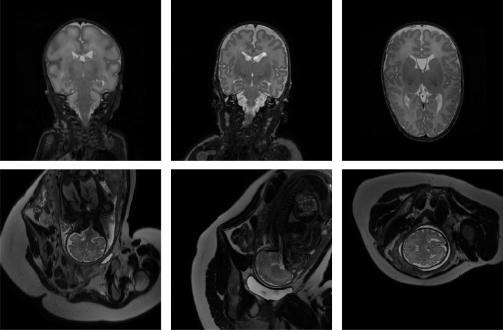
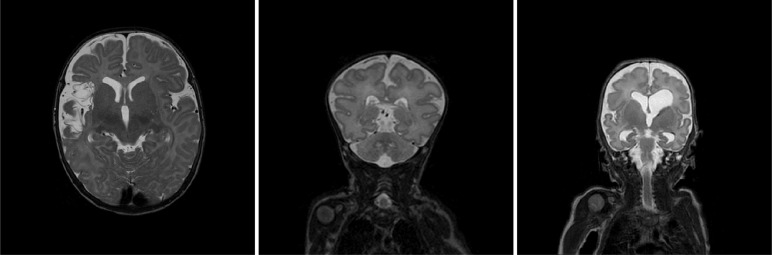
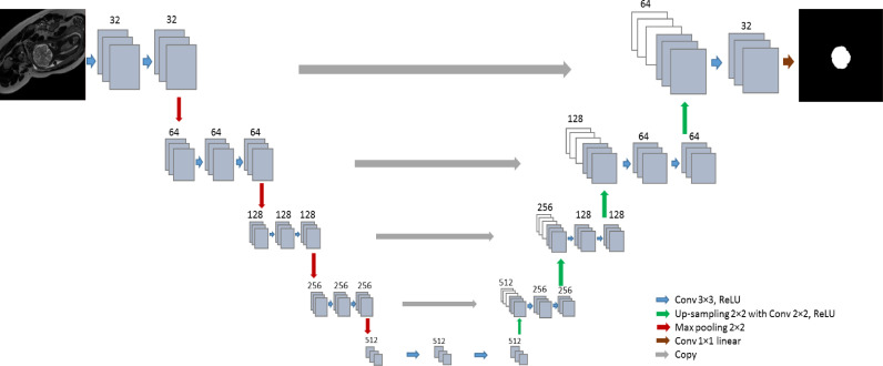
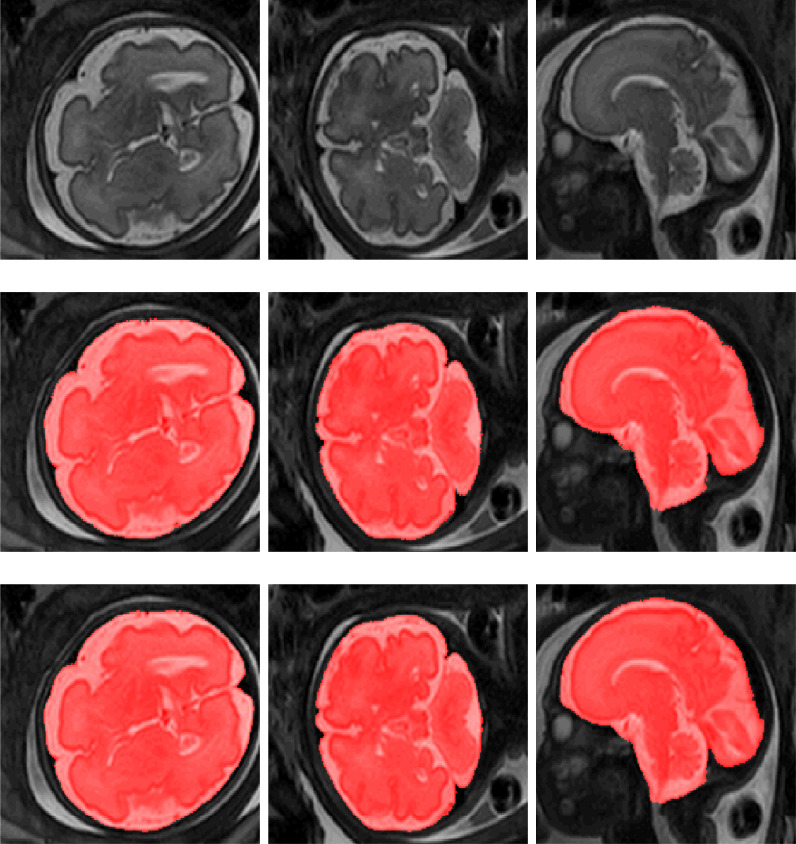
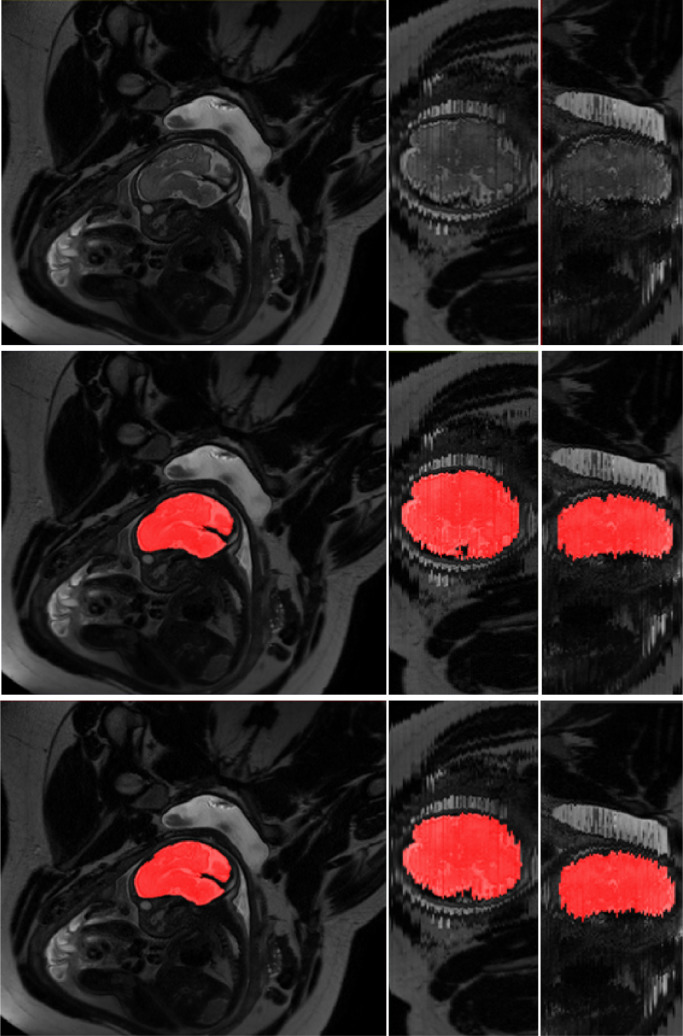
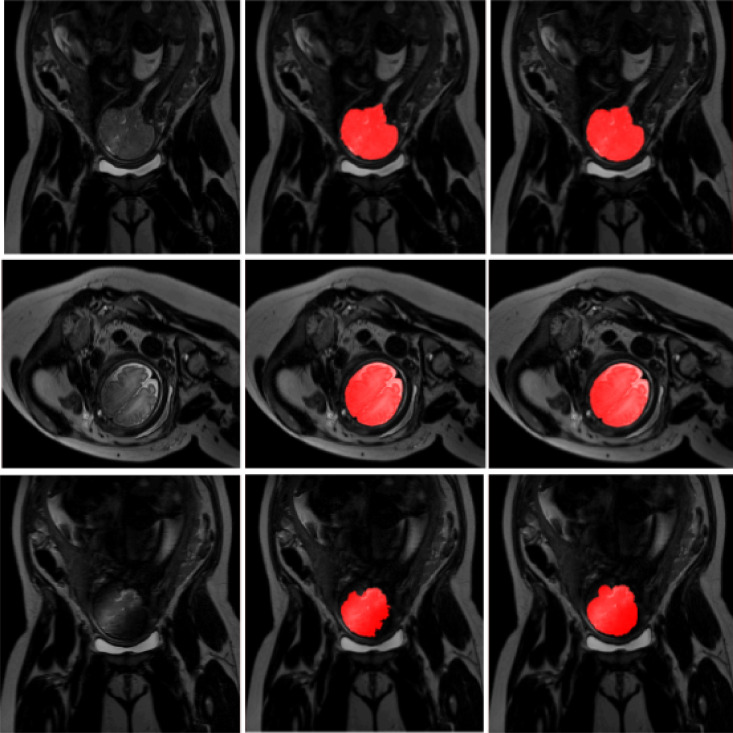
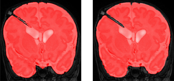
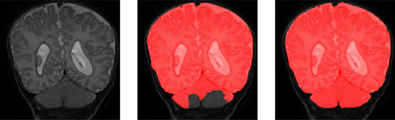
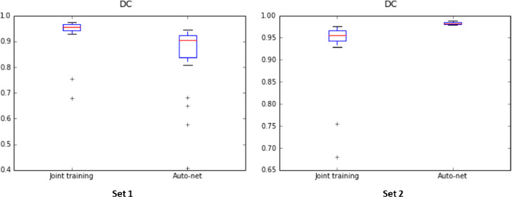
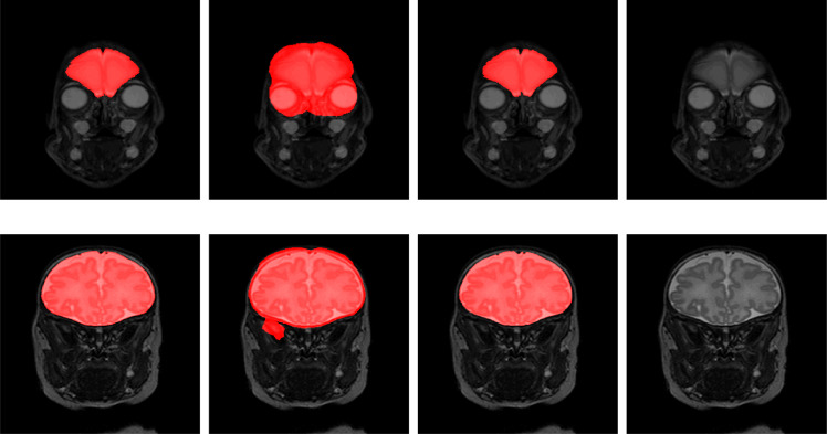
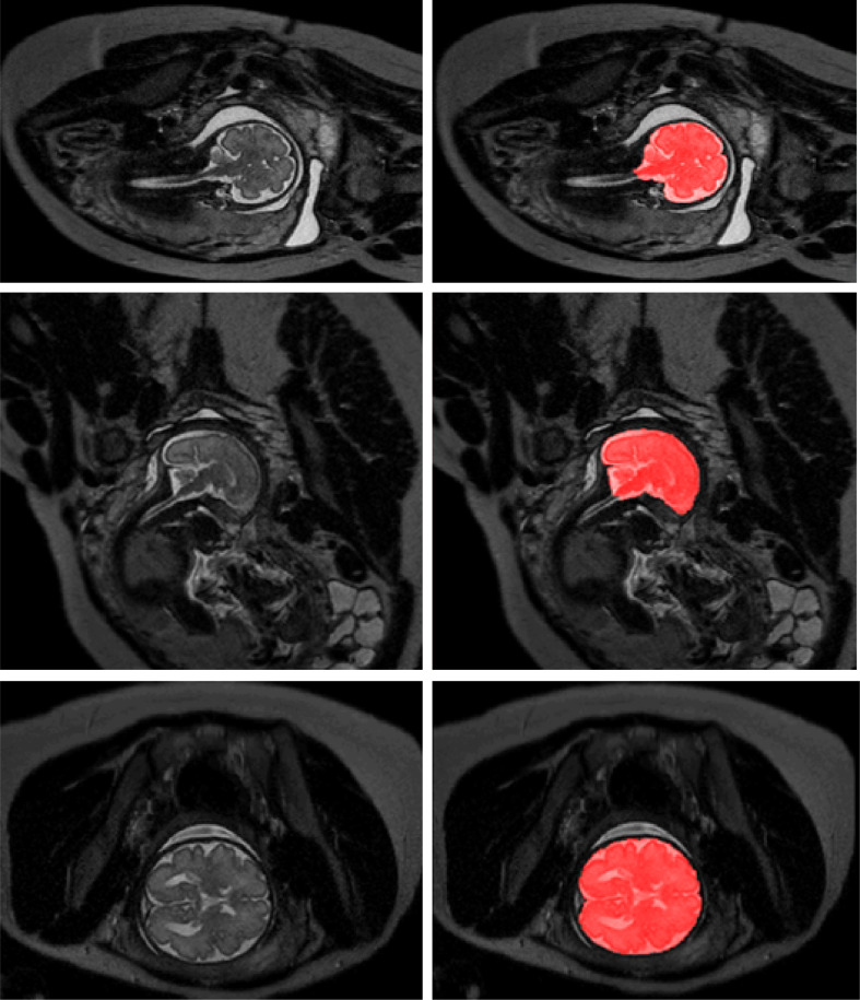
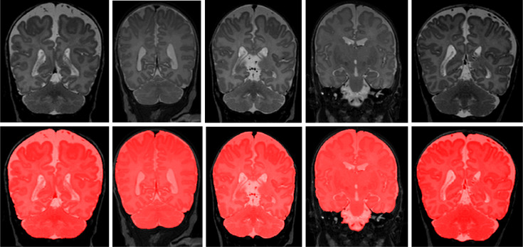
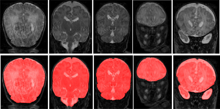
MR images of infants and fetuses allow non-invasive analysis of the brain. Quantitative analysis of brain development requires automatic brain tissue segmentation that is typically preceded by segmentation of the intracranial volume (ICV). Fast changes in the size and morphology of the developing brain, motion artifacts, and large variation in the field of view make ICV segmentation a challenging task. We propose an automatic method for segmentation of the ICV in fetal and neonatal MRI scans. The method was developed and tested with a diverse set of scans regarding image acquisition parameters (i.e. field strength, image acquisition plane, image resolution), infant age (23-45 weeks post menstrual age), and pathology (posthaemorrhagic ventricular dilatation, stroke, asphyxia, and Down syndrome). The results demonstrate that the method achieves accurate segmentation with a Dice coefficient (DC) ranging from 0.98 to 0.99 in neonatal and fetal scans regardless of image acquisition parameters or patient characteristics. Hence, the algorithm provides a generic tool for segmentation of the ICV that may be used as a preprocessing step for brain tissue segmentation in fetal and neonatal brain MR scans.

Keywords: Brain extraction; Brain segmentation; Deep learning; Fetal MRI; Intracranial volume segmentation; Neonatal MRI; Skull stripping.


Conflict of interest statement

There is no conflict between authors.

Figures

Fig. 1
Fig. 1
Examples of preterm neonatal and fetal MR scans included in the study. Top: coronal MRI acquired at 30 weeks PMA (left), coronal MRI acquired at 40 weeks PMA (middle), axial MRI acquired at 40 weeks PMA (right). Bottom: fetal MRI acquired in coronal (left), sagittal (middle) and axial (right) directions.
Fig. 2
Fig. 2
Examples of T2-weighted MR scans of preterm born neonates with ischemic stroke (left), Down syndrome (middle), and PHVD (right).
Fig. 3
Fig. 3
Network architecture: The network consists of a contracting path and an expanding path. The contracting path consists of repeated convolution layers followed by max pooling, and the expansion path consists of convolution layers followed by up-sampling.
Fig. 4
Fig. 4
Examples of ICV segmentation in slices from fetal scans acquired in axial (left), coronal (middle) and sagittal (right) image planes. The images are selected from the test set. A slice from T2-weighted image (top); segmentation achieved by the proposed method trained with a combination of neonatal and fetal MRI (middle); manual segmentation (bottom).
Fig. 5
Fig. 5
Examples of ICV segmentation in a scan acquired in sagittal plane. A slice from T2-weighted fetal MRI scan (top row), segmentation obtained with joint training (middle row) and manual segmentation (bottom row). The first column illustrates the segmentation in the in-plane view. Second and third columns illustrated out-of-plane views. The slices were selected from the test set.
Fig. 6
Fig. 6
Examples of ICV segmentation in slices from fetal scans that visualized intensity inhomogeneity. A slice from T2-weighted image (left); segmentation achieved by the proposed method trained with a combination of neonatal and fetal MRI (middle) and manual segmentation (right). The images were selected from the test set.
Fig. 7
Fig. 7
Example of ICV segmentation in one test neonate with PHVD on the left compared with manual segmentation on the right.The infants received a temporary ventricular shunt that is visible in some slices. The images were selected from the test set.
Fig. 8
Fig. 8
A slice from a scan of infant with PHVD (left) where the joint training undersegmented cerebellum (middle) compared with reference annotation (right). The cerebellar volume, shape and image intensity are typically different in infants with PHVD from infants without visible pathology. The images were selected from the test set.
Fig. 9
Fig. 9
Dice coefficients achieved by the proposed method using joint training with 21 fetal and 9 neonatal MRI scans, and by the publicly provided Auto-net trained with 260 fetal MRI scans. Both methods were tested on Set 1 (left) and Set 2 (right).
Fig. 10
Fig. 10
Examples of ICV segmentation in neonates acquired at 30 weeks PMA (top row) and 40 weeks PMA (bottom row). Results of the joint training (first column), the result obtained with BET (second column), manual annotation (third column) and the original T2-weighted MRI (last column).The images were selected from the test set.
Fig. 11
Fig. 11
Examples of ICV segmentation in slices from fetal scans acquired with 1.5 Tesla scanner in coronal (top), sagittal (middle) and axial (bottom) image planes. A slice from T2-weighted image (left) and segmentation achieved by the proposed method trained with a combination of neonatal and fetal MRIs (right).
Fig. 12
Fig. 12
Examples of ICV segmentation in 5 neonatal MR scans with intensity inhomogeneity artifacts. A slice from T2-weighted fetal MRI scan (first row); segmentation obtained with joint training (second row).
Fig. 13
Fig. 13
Examples of ICV segmentation in 5 neonatal MR scans with motion artifacts. A slice from T2-weighted fetal MRI scan (first row); segmentation obtained with joint training (second row).

References

    1. Akkus Z., Galimzianova A., Hoogi A., Rubin D.L., Erickson B.J. Deep learning for brain MRI segmentation: state of the art and future directions. J. Digit. Imaging. 2017;30(4):449–459. - PMC - PubMed
    1. Alderliesten T., de Vries L.S., Staats L., van Haastert I.C., Weeke L., Benders M.J., Koopman-Esseboom C., Groenendaal F. MRI and spectroscopy in (near) term neonates with perinatal asphyxia and therapeutic hypothermia. Arch. Dis. Child. Fetal Neonatal Edition. 2017;102(2):F147–F152. - PubMed
    1. Anquez J., Angelini E.D., Bloch I. The IEEE International Symposium on Biomedical Imaging (ISBI) IEEE; 2009. Automatic segmentation of head structures on fetal MRI; pp. 109–112.
    1. Benders M.J., van der Aa N.E., Roks M., van Straaten H.L., Isgum I., Viergever M.A., Groenendaal F., de Vries L.S., van Bel F. Feasibility and safety of erythropoietin for neuroprotection after perinatal arterial ischemic stroke. J. Pediatr. 2014;164(3):481–486. - PubMed
    1. Brouwer M.J., De Vries L.S., Kersbergen K.J., Van Der Aa N.E., Brouwer A.J., Viergever M.A., Išgum I., Han K.S., Groenendaal F., Benders M.J. Effects of posthemorrhagic ventricular dilatation in the preterm infant on brain volumes and white matter diffusion variables at term-equivalent age. J. Pediatr. 2016;168:41–49. - PubMed

Publication types