

Cell-Electrospinning and Its Application for Tissue Engineering
- PMID: 31835356
- PMCID: PMC6940787
- DOI: 10.3390/ijms20246208
Cell-Electrospinning and Its Application for Tissue Engineering
Abstract
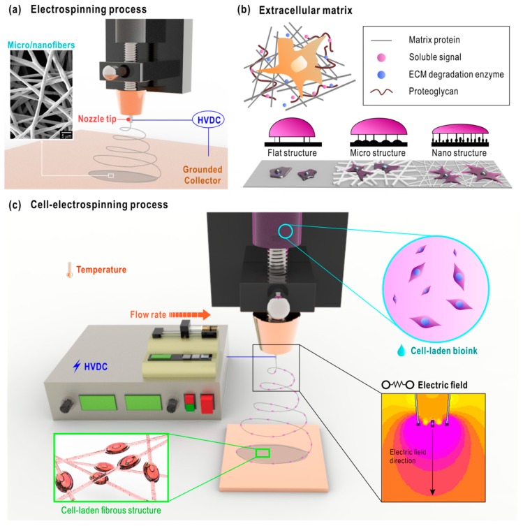
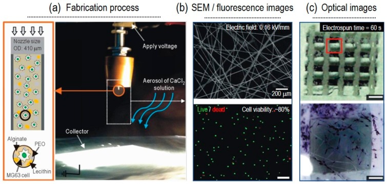
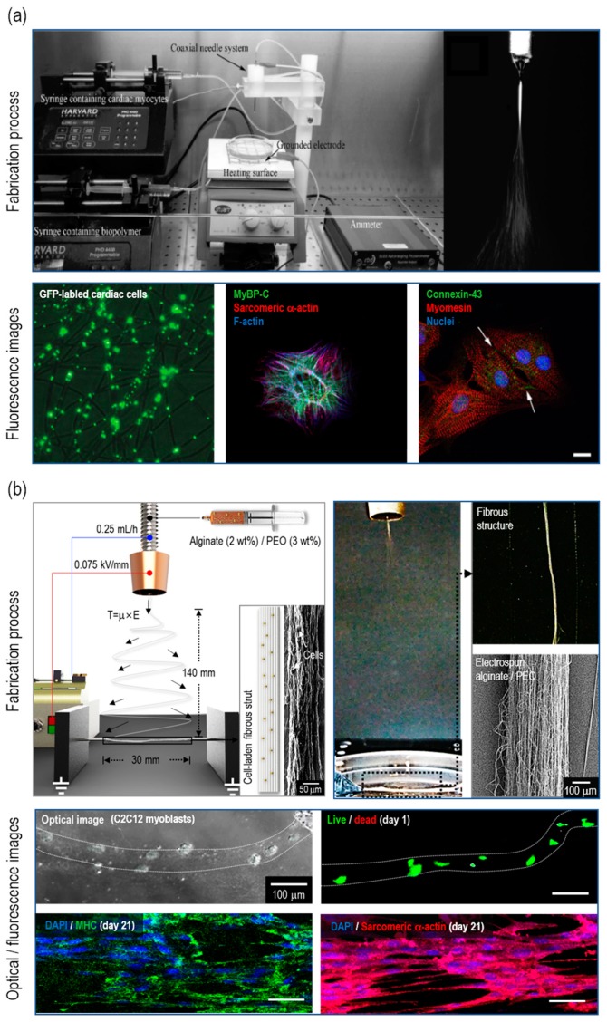
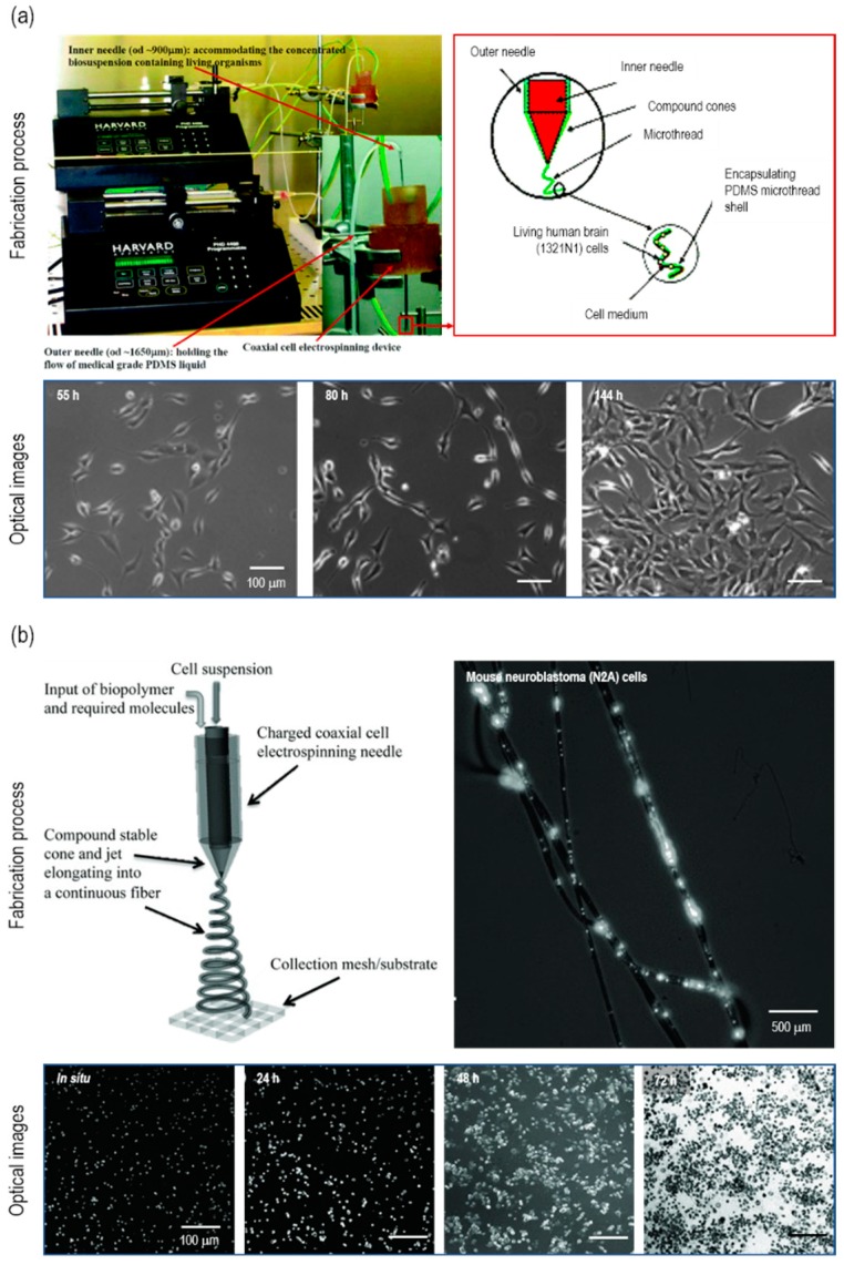
Electrospinning has gained great interest in the field of regenerative medicine, due to its fabrication of a native extracellular matrix-mimicking environment. The micro/nanofibers generated through this process provide cell-friendly surroundings which promote cellular activities. Despite these benefits of electrospinning, a process was introduced to overcome the limitations of electrospinning. Cell-electrospinning is based on the basic process of electrospinning for producing viable cells encapsulated in the micro/nanofibers. In this review, the process of cell-electrospinning and the materials used in this process will be discussed. This review will also discuss the applications of cell-electrospun structures in tissue engineering. Finally, the advantages, limitations, and future perspectives will be discussed.
Keywords: cell-electrospinning; cell-laden scaffold; micro/nano structure; tissue engineering.
Conflict of interest statement
The authors declare no conflict of interest.
Figures
References
-
- Tucker N., Stanger J.J., Staiger M.P., Razzaq H., Hofman K. The history of the science and technology of electrospinning from 1600 to 1995. J. Eng. Fiber Fabr. 2012;7:155892501200702S10. doi: 10.1177/155892501200702S10. - DOI
-
- Mercante L.A., Scagion V.P., Migliorini F.L., Mattoso L.H., Correa D.S. Electrospinning-based (bio) sensors for food and agricultural applications: A review. Trends Anal. Chem. 2017;91:91–103. doi: 10.1016/j.trac.2017.04.004. - DOI
-
- Nisha M., Singh D., Freesta Shiny J., Sasirekha B. Design and Manufacture of Nanofibers Using Electrospinning Technique for Aerospace Application. Appl. Mech. Mater. 2016;852:72–78. doi: 10.4028/www.scientific.net/AMM.852.72. - DOI
-
- Dotti F., Varesano A., Montarsolo A., Aluigi A., Tonin C., Mazzuchetti G. Electrospun porous mats for high efficiency filtration. J. Ind. Textil. 2007;37:151–162. doi: 10.1177/1528083707078133. - DOI
Publication types
MeSH terms
Grants and funding
LinkOut - more resources
Full Text Sources
