. 2019 Dec 10;9(12):851.
doi: 10.3390/biom9120851.

Alcohol Metabolism Potentiates HIV-Induced Hepatotoxicity: Contribution to End-Stage Liver Disease

Affiliations

Alcohol Metabolism Potentiates HIV-Induced Hepatotoxicity: Contribution to End-Stage Liver Disease

Murali Ganesan et al. Biomolecules. .

Abstract

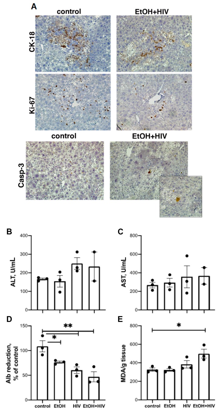
In an era of improved survival due to modern antiretroviral therapy, liver disease has become a major cause of morbidity and mortality, resulting in death in 15-17% of human immunodeficiency virus (HIV)-infected patients. Alcohol enhances HIV-mediated liver damage and promotes the progression to advanced fibrosis and cirrhosis. However, the mechanisms behind these events are uncertain. Here, we hypothesize that ethanol metabolism potentiates accumulation of HIV in hepatocytes, causing oxidative stress and intensive apoptotic cell death. Engulfment of HIV-containing apoptotic hepatocytes by non-parenchymal cells (NPCs) triggers their activation and liver injury progression. This study was performed on primary human hepatocytes and Huh7.5-CYP cells infected with HIV-1ADA, and major findings were confirmed by pilot data obtained on ethanol-fed HIV-injected chimeric mice with humanized livers. We demonstrated that ethanol exposure potentiates HIV accumulation in hepatocytes by suppressing HIV degradation by lysosomes and proteasomes. This leads to increased oxidative stress and hepatocyte apoptosis. Exposure of HIV-infected apoptotic hepatocytes to NPCs activates the inflammasome in macrophages and pro-fibrotic genes in hepatic stellate cells. We conclude that while HIV and ethanol metabolism-triggered apoptosis clears up HIV-infected hepatocytes, continued generation of HIV-expressing apoptotic bodies may be detrimental for progression of liver inflammation and fibrosis due to constant activation of NPCs.

Keywords: acetaldehyde; apoptotic bodies; ethanol; fibrosis; hepatocytes; human immunodeficiency virus; inflammation.


Conflict of interest statement

The authors declare no conflict of interest.

Figures

Figure 1
Figure 1
Human immunodefeciency virus (HIV) and ethanol induce apoptosis in hepatocytes: Data are from three independent experiments presented as means ±standard error, (SE). Bars marked with the same letter are not significantly different from each other; bars with different letters are significantly different (p ≤ 0.05). (A) Effects of various EtOH treatment regimens on caspase 3 cleavage in HIV-exposed hepatocytes (Western blotting, WB). (B) Prolonged expression of alcohol-metabolizing enzymes, CYP2E1 and ADH, in hepatocytes plated on polyelectrolyte multilayer (PEM) gels. We cut a unnecessary group in third lane, so we joined the last lane. (C) M30 ELISA (apoptosis) in HIV-infected hepatocytes in the presence or absence of azidothymidine (AZT) (100 µM). (D) Effects of various HIV MOI on caspase 3 cleavage in RLW cells treated with an acetaldehyde-generating system (AGS).
Figure 2
Figure 2
Effects of ethanol metabolism on human immunodeficiency virus gag ribonuclear acid (HIVgag RNA) expression in HIV-infected hepatocytes. All data are from three independent experiments presented as means ± SE. Bars marked with the same letter are not significantly different from each other; bars with different letters are significantly different (p ≤ 0.05). (A) Effects of pancaspase inhibitor (PCI) on HIV gag RNA in primary human hepatocytes. (B) Effects of (AZT and 4-methyl pyrazole (4MP) on HIV RNA in primary human hepatocytes. (C) Reverse transcriptase (RT) activity in supernatants of primary human hepatocytes. (D) RT activity in supernatants of RLW cells exposed to AGS. (E) The HIVgag RNA after removal of membrane-expressed structures by low acid wash in RLW cells.
Figure 3
Figure 3
Effect of acetaldehyde generating system (AGS) on HIV-DNA levels in RLW cells. The results from duplicated representative experiment are shown in panels (A,B). (A) Effects of PCI on intracellular HIV DNA. (B) AGS increased the level of integrated HIV DNA (in the presence of PCI).
Figure 4
Figure 4
Kinetics of HIV components, oxidative stress markers and cleaved caspase-3 in HIV-infected RLW cells exposed to AGS. Data are from three independent experiments presented as means ± SE. Bars marked with the same letter are not significantly different from each other; bars with different letters are significantly different (p ≤ 0.05). (A) HIVgag RNA; (B) p24; (C) Cleaved caspase-3; (D) reactive oxygen species (ROS); (E) 4-hydroxynonenal ( 4HNE) protein adducts
Figure 5
Figure 5
Effects of AGS on possible receptors for viral entry on hepatocytes (the results of representative experiments). (A) C-X-C-chemokine receptor type 4 CXCR4; (B) C-C-chemokine receptor type 5 (CCR5); (C) Galactosyl ceramide (GalCer).
Figure 6
Figure 6
The AGS exposure promotes accumulation of HIV components by suppressing lysosome and proteasome activities in RLW cells: Data are from three independent experiments presented as means ± SE. Bars marked with the same letter are not significantly different from each other; bars with different letters are significantly different (p ≤ 0.05). (A) Chloroquine enhances expression of HIV gag RNA; (B) Lysosome and proteasome inhibitors stabilize p24 expression; (C) Chymotrypsin-like proteasome activity; (D) Trypsin-like proteasome activity; (E) Cathepsin L-activity; (F) Cathepsin B activity.
Figure 7
Figure 7
Gene activation in hepatocytes exposed to HIV and EtOH. Data are from three independent experiments presented as means ± SE. Bars marked with the same letter are not significantly different from each other; bars with different letters are significantly different (p ≤ 0.05): (A) Next-generation sequencing (NGS, heatmap); (B) Expression of HIV genes (NGS); (C) Activation of interferon-stimulated genes (ISGs) in RLW cells. (D) Activation of pro-apoptotic genes in RLW cells.
Figure 8
Figure 8
Activation of monocyte-derived macrophages (MDMs) and hepatic stellate cells (HSCs) by engulfment of ABHep. Data are from three independent experiments presented as means ± SE. Bars marked with the same letter are not significantly different from each other; bars with different letters are significantly different (p ≤ 0.05). (A) Inflammasome markers in MDMs. (B) Pro-fibrotic gene activation in HSCs. (C) Inflammasome markers in mRNA expression in lymphocytes. (D) Pro-fibrotic mRNAs in lymphocytes.
Figure 9
Figure 9
Ethanol feeding depletes human hepatocytes in HIV-exposed mice with humanized livers (pilot study). (A) Liver morphology (mice fed control diet vs. mice fed ethanol diet, all injected with HIV). Liver tissue was stained with the human-specific antibodies cytokeratin (CK)-18 and KI-67 as well as with anti-caspase-3. (B) alanine aminotransferase ALT. (C) aspartate aminotransferase (AST). (D) Human albumin (reduction, % control). (E) thiobarbituric acid reactive substances (TBARS).
Figure 10
Figure 10
Ethanol metabolism promotes apoptosis in HIV-infected hepatocytes, causing liver inflammation and fibrosis development. Ethanol metabolism suppresses lysosome and proteasome activities in HIV-infected hepatocytes and causes accumulation of HIV components, finally leading to apoptosis induction and abortive HIV replication. Apoptotic hepatocytes that contain viral components are engulfed by macrophages (MDMs) and HSCs, thereby promoting activation of inflammasome in MDMs and activation of pro-fibrotic genes in HSCs, leading to liver inflammation and fibrosis development.

References

    1. Debes J.D., Bohjanen P.R., Boonstra A. Mechanisms of accelerated liver fibrosis progression during hiv infection. J. Clin. Transl. Hepatol. 2016;4:328–335. - PMC - PubMed
    1. Pascual-Pareja J.F., Caminoa A., Larrauri C., Gonzalez-Garcia J., Montes M.L., Diez J., Grande M., Arribas J.R. Haart is associated with lower hepatic necroinflammatory activity in hiv-hepatitis c virus-coinfected patients with cd4 cell count of more than 350 cells/microl at the time of liver biopsy. AIDS. 2009;23:971–975. doi: 10.1097/QAD.0b013e328329f994. - DOI - PubMed
    1. Mata-Marin J.A., Gaytan-Martinez J., Grados-Chavarria B.H., Fuentes-Allen J.L., Arroyo-Anduiza C.I., Alfaro-Mejia A. Correlation between hiv viral load and aminotransferases as liver damage markers in hiv infected naive patients: A concordance cross-sectional study. Virol. J. 2009;6:181. doi: 10.1186/1743-422X-6-181. - DOI - PMC - PubMed
    1. Penton P.K., Blackard J.T. Analysis of hiv quasispecies suggests compartmentalization in the liver. AIDS Res. Hum. Retrovir. 2014;30:394–402. doi: 10.1089/aid.2013.0146. - DOI - PMC - PubMed
    1. Sterling R.K., Chiu S., Snider K., Nixon D. The prevalence and risk factors for abnormal liver enzymes in hiv-positive patients without hepatitis B or C coinfections. Dig. Dis. Sci. 2008;53:1375–1382. doi: 10.1007/s10620-007-9999-6. - DOI - PMC - PubMed

Publication types

MeSH terms

LinkOut - more resources