. 2019 Dec 10;20(24):6234.
doi: 10.3390/ijms20246234.

Effect of Methionine Diet on Metabolic and Histopathological Changes of Rat Hippocampus

Affiliations

Effect of Methionine Diet on Metabolic and Histopathological Changes of Rat Hippocampus

Maria Kovalska et al. Int J Mol Sci. .

Abstract

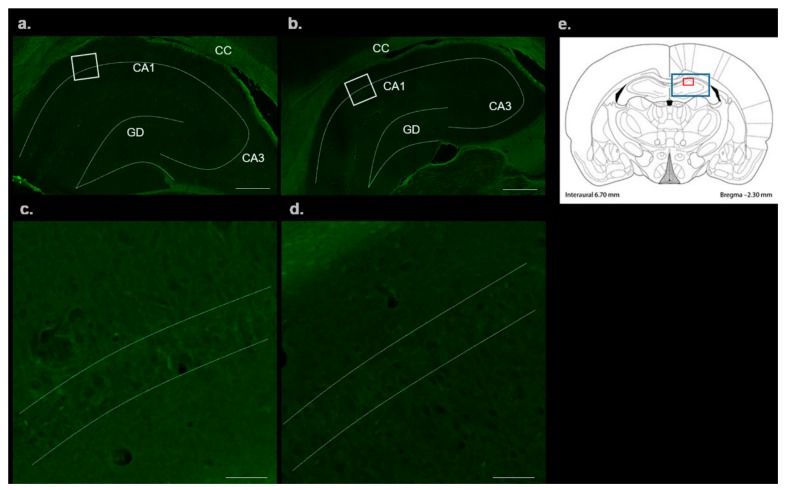
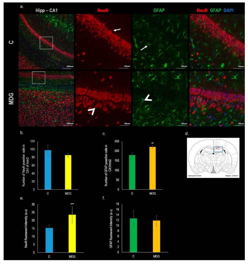
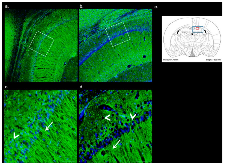
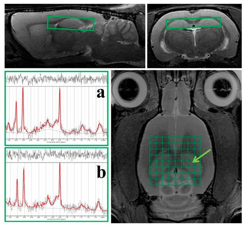
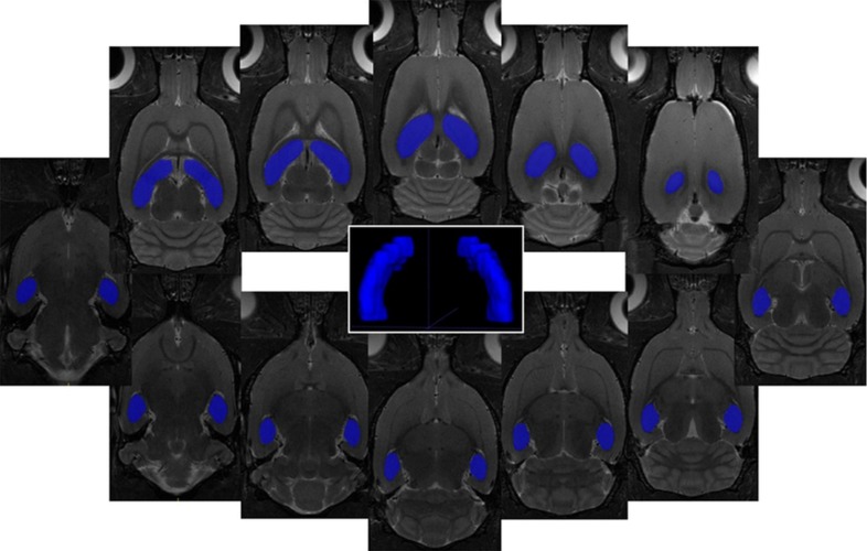
Hyperhomocysteinemia (hHcy) is regarded as an independent and strong risk factor for cerebrovascular diseases, stroke, and dementias. The hippocampus has a crucial role in spatial navigation and memory processes and is being constantly studied for neurodegenerative disorders. We used a moderate methionine (Met) diet at a dose of 2 g/kg of animal weight/day in duration of four weeks to induce mild hHcy in adult male Wistar rats. A novel approach has been used to explore the hippocampal metabolic changes using proton magnetic resonance spectroscopy (1H MRS), involving a 7T MR scanner in combination with histochemical and immunofluorescence analysis. We found alterations in the metabolic profile, as well as remarkable histo-morphological changes such as an increase of hippocampal volume, alterations in number and morphology of astrocytes, neurons, and their processes in the selective vulnerable brain area of animals treated with a Met-enriched diet. Results of both methodologies suggest that the mild hHcy induced by Met-enriched diet alters volume, histo-morphological pattern, and metabolic profile of hippocampal brain area, which might eventually endorse the neurodegenerative processes.

Keywords: 1H MRS; hippocampus; hyperhomocysteinemia; methionine diet; neurodegeneration.


Conflict of interest statement

The authors declare no conflict of interest.

Figures

Figure 1
Figure 1
FluoroJade-C (FJC) stained rat brain sections in the cornu ammonis 1 (CA1) region of hippocampus. Fluorescent micrographs of rat hippocampus representing control (a) and MDG (b) with a detail of corresponding group focusing on CA1 region of control (c) and MDG group (d). White square in the first column presents area of magnification. (a,b) Bar = 500 μm; (c,d) Bar = 50 μm; n = 5/group. Schematic coronal rat brain section (e), redrawn according to Tothova et al. [12] representing hippocampus (blue rectangle) and smaller (red rectangle) detects CA1 area of rat hippocampus.
Figure 2
Figure 2
Immunofluorescent microphotographs and statistical evaluation of NeuN and GFAP in the CA1 region of hippocampus. (a) Fluorescent micrographs of rat hippocampus representing control and Met-enriched diet treated animals (MDG). The first column represents low magnification pictures of hippocampus in CA1 region of both experimental groups. The second column determines high magnification pictures of CA1 stained by NeuN (red). The third column indicates high magnification pictures of CA1 stained by glial fibrillary acidic protein (GFAP) (green) and the last column portrays high magnification pictures of CA1 of overlaid antibodies contra-stained by 4′,6-diamidino-2-phenylindole (DAPI; blue). The white rectangle in the first column presents the area of magnification. Arrows point to neurons and astrocytes without any impairment. Arrowheads indicate morphologically changed neurons or astrocytes. (a) (column Hipp-CA1) Bar = 200 μm; (a) (columns NeuN, GRAF and NeuN + GFAP + DAPI) = 50 μm; n = 5/group. Schematic coronal rat brain section (d), described in Figure 1. (b) Number of NeuN positive neurons in the CA1 region in the control group (C) and MDG group. (c) Number of GFAP positive astrocytes in the CA1 region. (e) NeuN fluorescent intensity in the CA1 region. (f) GFAP fluorescent intensity in the CA1 region. All results are presented as mean ± SD for n = 5/group, normalized to the control levels. *** p < 0.001 and ** p < 0.01 versus the control value.
Figure 3
Figure 3
Immunofluorescent microphotographs of β-Tubulin in the CA1 region of hippocampus. Fluorescent micrographs of low magnification of rat CA1 of hippocampus representing control (a) and Met-enriched diet treated animals (b). The second row represents details of the corresponding group focusing on the CA1 region of hippocampus ((c): C and (d): MDG). Arrows indicate axons, arrowheads body on neurons. Nuclei are contra-stained by DAPI (blue). The white square in the first column presents area of magnification. Schematic coronal rat brain section (e), described in Figure 1. (a,b) Bar = 200 μm; (c,d) Bar = 50 μm; n = 5/group.
Figure 4
Figure 4
Representative picture of in vivo 1H MRS in the hippocampus of the rat brain. On the morphological T2-weighted MR images is shown the position of the CSI grid with real voxel size of 2.75 × 2.75 × 2 mm3 covering the hippocampus (green grid and boxes on MRI) of the rat brain. There are also displayed representative in vivo 1H MRS-spectra from the selected voxel (green arrow) in the rat hippocampus from control (a) and MDG groups (b) evaluated in LCModel software (version 6.3-1K; S. Provencher, Oakville, ON, Canada).
Figure 5
Figure 5
Representative pictures of in vivo MRI volumetry of the hippocampus of the rat brain. On 12 consecutive coronal T2-weighted MRI (resolution 0.137 × 0.137 × 0.5 mm3) is displayed representative ROIs covering the hippocampus of the rat brain together with 3D visualization of target areas plotted by ITK-SNAP software (Version 3.4.0, US National Institutes of Health, Philadelphia, PA, USA).

References

    1. Cardoso B.R., Cominetti C., Cozzolino S.M. Importance and management of micronutrient deficiencies in patients with Alzheimer’s disease. Clin. Interv. Aging. 2013;8:531–542. doi: 10.2147/CIA.S27983. - DOI - PMC - PubMed
    1. Berti V., Murray J., Davies M., Spector N., Tsui W.H., Li Y., Williams S., Pirraglia E., Vallabhajosula S., McHugh P., et al. Nutrient patterns and brain biomarkers of Alzheimer’s disease in cognitively normal individuals. J. Nutr. Health Aging. 2015;19:413–423. doi: 10.1007/s12603-014-0534-0. - DOI - PMC - PubMed
    1. Xu Y., Tian Y., Wei H.J., Dong J.F., Zhang J.N. Methionine diet-induced hyperhomocysteinemia accelerates cerebral aneurysm formation in rats. Neurosci. Lett. 2011;494:139–144. doi: 10.1016/j.neulet.2011.02.076. - DOI - PubMed
    1. Liu B., Ma S., Wang T., Zhao C., Li Y., Yin J., Liu C., Gao C., Sun L., Yue W., et al. A novel rat model of heart failure induced by high methionine diet showing evidence of association between hyperhomocysteinemia and activation of NF-kappaB. Am. J. Transl. Res. 2016;8:117–124. - PMC - PubMed
    1. Esse R., Barroso M., de Almeida I.T., Castro R. The Contribution of Homocysteine Metabolism Disruption to Endothelial Dysfunction: State-of-the-Art. Int. J. Mol. Sci. 2019;20:867. doi: 10.3390/ijms20040867. - DOI - PMC - PubMed

LinkOut - more resources