. 2019 Dec 11;8(12):1606.
doi: 10.3390/cells8121606.

Maternal Yes-Associated Protein Participates in Porcine Blastocyst Development via Modulation of Trophectoderm Epithelium Barrier Function

Affiliations

Maternal Yes-Associated Protein Participates in Porcine Blastocyst Development via Modulation of Trophectoderm Epithelium Barrier Function

Zubing Cao et al. Cells. .

Abstract

The establishment of a functional trophectoderm (TE) epithelium is an essential prerequisite for blastocyst formation and placentation. Transcription coactivator yes-associated protein (YAP), a downstream effector of the hippo signaling pathway, is required for specification of both the TE and epiblast lineages in mice. However, the biological role of YAP in porcine blastocyst development is not known. Here, we report that maternally derived YAP protein is localized to both the cytoplasm and nuclei prior to the morula stage and is then predominantly localized to the TE nuclei in blastocysts. Functionally, maternal YAP knockdown severely impeded blastocyst formation and perturbed the allocation of the first two lineages. The treatment of embryos with verteporfin, a pharmacological inhibitor of YAP, faithfully recapitulated the phenotype observed in YAP deleted embryos. Mechanistically, we found that maternal YAP regulates multiple genes which are important for lineage commitment, tight junction assembly, and fluid accumulation. Consistent with the effects on tight junction gene expression, a permeability assay revealed that paracellular sealing was defective in the trophectoderm epithelium. Lastly, YAP knockdown in a single blastomere at the 2-cell stage revealed that the cellular progeny of the YAP+ blastomere were sufficient to sustain blastocyst formation via direct complementation of the defective trophectoderm epithelium. In summary, these findings demonstrate that maternal YAP facilitates porcine blastocyst development through transcriptional regulation of key genes that are essential for lineage commitment, tight junction assembly, and fluid accumulation.

Keywords: YAP; blastocyst development; pig; tight junction; trophectoderm.


Conflict of interest statement

The authors declare no conflict of interest.

Figures
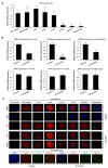
Figure 1
Figure 1
Expression of maternal YAP mRNA and protein in porcine early embryos. (A) Expression of YAP mRNA in oocytes and early embryos. Relative abundance of YAP mRNA was determined by qPCR. Data were normalized against endogenous reference gene EF1α1 and the data from each stage were relative to GV oocyte. Data are shown as mean ± S.E.M and different letters on the bars indicate significant differences (p < 0.05). (B) Expression of TEAD4 and YAP mRNA in embryos treated with or without α-amanitin. Relative abundance of TEAD4 and YAP mRNA in 4-cell, 8-cell, and blastocysts was determined by qPCR. Data were normalized against endogenous reference gene EF1α1 and the data from each stage were relative to the control group. Data are shown as mean ± S.E.M and different letters on the bars indicate significant differences (p < 0.05). (C) Expression and localization of YAP in oocytes and early embryos derived from Parthenogenetic Activation (PA) or In Vitro Fertilization (IVF). Oocytes and PA embryos at the indicated stages were stained for YAP (red) and DNA (blue). IVF blastocysts were double stained for YAP (red) and SOX2 (green). Representative Z-stack and Z-section images obtained by confocal microscopy are shown. The experiment was independently repeated three times with at least 20 oocytes or embryos per stage. Scale bar: 50 µm.
Figure 2
Figure 2
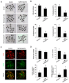
Validation of YAP knockdown efficiency in porcine embryos. (A) Expression levels of YAP mRNA in embryos. The relative abundance of YAP mRNA in 2-cell, 4-cell, and 8-cell embryos from control, sham water injection, and siRNA injection was determined by qPCR. Data were normalized against endogenous reference gene EF1α1 and the data from each stage were relative to the control group. Data are shown as mean ± S.E.M and different letters on the bars indicate significant differences (p < 0.05). (B) Expression and localization of YAP protein in embryos. Two-cell, 4-cell, and morula stage embryos from each group were stained to indicate YAP (red) and DNA (blue). Representative images obtained by confocal microscopy are shown. The experiment was independently repeated three times with at least 24 embryos per stage. Scale bar: 50 µm. (C) Western blot analysis of YAP protein expression. Four-cell embryos from each group were used for western blot analysis and α-TUBULIN was used as a loading control. A representative image is shown. (D) Quantitative analysis of YAP protein expression using western blot. Data are expressed as mean ± S.E.M from three independent experiments and different letters on the bars indicate significant differences (p < 0.05).
Figure 3
Figure 3
Effect of YAP knockdown on the developmental efficiency of porcine embryos. (A) Representative images of embryos at different stages. MII oocytes were microinjected with YAP siRNA. Uninjected oocytes or sham injected (water) served as two control groups. MII oocytes from each group were then parthenogenetically activated and cultured to the blastocyst stage. Scale bar: 100 µm. (B) Developmental rates of porcine preimplantation embryos. The rates of 8-cell embryos and blastocysts at day 5, 6, and 7 were recorded and statistically analyzed in each group. Data are expressed as mean ± S.E.M and different letters on the bars indicate significant differences (p < 0.05). (C) Immunofluorescence staining of blastocysts in each group using a CDX2 antibody. Blastocysts were stained to indicate CDX2 (green) and DNA (red). Representative images obtained using confocal microscopy are shown. The experiment was independently repeated three times with at least 10 blastocysts per group. The bottom panel in each group shows merged images between CDX2 and DNA. Scale bar: 50 µm. (D) Lineage allocation analysis of YAP knockdown and control blastocysts. Total cell numbers, TE cells, CDX2 negative cells, and the ratio of CDX2 negative cells to TE cells were separately recorded and subjected to statistical analysis. TE: trophectoderm. Data are represented as mean ± S.E.M and different letters on the bars indicate significant differences (p < 0.05).
Figure 4
Figure 4
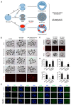
Effect of YAP inhibition on the developmental efficiency of porcine embryos. (A) Representative images of embryos at different stages from control and verteporfin treatment groups. One-cell embryos were cultured in vitro for 7 days in the presence of 1 μM verteporfin (YAP inhibitor) dissolved in DMSO. Embryos cultured in medium containing an equivalent amount of DMSO served as a control group. Scale bar: 100 µm. (B) The developmental rates of early embryos cultured with or without verteporfin. Developmental rates of 8-cell embryos and blastocysts on day 5, 6, and 7 were recorded in each group. Data are expressed as mean ± S.E.M and different letters denote significant differences (p < 0.05). (C) Representative fluorescence images of blastocysts stained with CDX2 antibody. Blastocysts were stained to indicate CDX2 (green) and DNA (red). The experiment was independently repeated three times with at least 10 blastocysts per group. The bottom panel in each group shows the merged images between CDX2 and DNA. Scale bar: 50 µm. (D) Lineage allocation analysis of YAP inhibited and control blastocysts. Total cell numbers, TE cells, CDX2 negative cells, and the ratio of CDX2 negative cells to TE cells were separately recorded and subjected to statistical analysis. TE: trophectoderm. Data are shown as mean ± S.E.M and different letters denote significant differences (p < 0.05).
Figure 5
Figure 5
YAP knockdown perturbs the expression of genes required for lineage commitment, TJ assembly, and fluid accumulation. (A) Expression of putative YAP target genes in control and YAP knockdown morula. Relative expression of YAP target genes was determined by qPCR. Data were normalized against an endogenous reference gene (EF1α1) and the data from the control were set to 1. Data are shown as mean ± S.E.M and different letters denote significant differences (p < 0.05). (B) Expression and localization of YAP target gene proteins in control and YAP knockdown morula. Target proteins and DNA are represented as green and red, respectively. Representative images obtained using confocal microscopy are shown. The experiment was independently repeated three times with at least 15 morula per group. Scale bar: 100 µm. (C) Representative brightfield and fluorescence images of FITC-dextran treated blastocysts from the control and YAP knockdown groups. Blastocysts in each group were incubated in the medium containing 1 mg/mL 40 kDa FITC-dextran for 30 min and then the blastocysts were visualized under an inverted fluorescence microscope. Scale bar: 100 µm. (D) Analysis of paracellular permeability in trophectoderm by FITC-dextran uptake assay. The number of FITC positive blastocysts in each group was statistically analyzed. Data are shown as mean ± S.E.M and different letters denote significant differences (p < 0.05).
Figure 6
Figure 6
YAP+ blastomeres complement YAP deleted blastomeres to sustain blastocyst development. (A) Experimental design describing YAP knockdown rescue experiments in embryos. PA: parthenogenetic activation, TE: trophectoderm. (B) Representative images of embryos at different stages from control, YAP knockdown in oocytes and YAP knockdown in single blastomere of 2-cell embryos. MII oocytes were microinjected with YAP siRNA. Single blastomere of a 2-cell embryo was co-microinjected with both YAP siRNA and mCherry mRNA. Uninjected MII oocytes served as a control. Embryos in each group were cultured until the blastocyst stage. The blastocysts were then visualized under an inverted fluorescence microscope. (C). Enlarged images of single blastocysts from each group is shown. Scale bar: 50 µm. (D) The developmental rates of early embryos. Proportion of embryos that developed to the 8-cell stage and blastocysts on day 5, 6, and 7 were recorded. Data are expressed as mean ± S.E.M and different letters denote significant differences (p < 0.05). (E) Expression and localization of both YAP and its target proteins in morula. Target proteins were evaluated using specific antibodies (green) and DNA was visualized using propidium iodide (red). Representative images obtained using confocal microscopy are shown. The experiment was independently repeated three times with at least 15 morula and blastocysts per group. Scale bar: 50 µm.
Figure 7
Figure 7
Working model illustrating how maternal YAP regulates trophectoderm integrity to facilitate porcine blastocyst development. In the TE epithelium, inactivation of hippo signaling induces the translocation of cytoplasmic YAP into the nucleus, which in turn binds to TEAD family proteins to form a transcriptional complex. The YAP-containing complex positively regulates the expression of genes (black) that are important for lineage commitment (CDX2, TEAD4, OCT4, and SOX2), TJ assembly (OCLN, CLDN4, CLDN6, CDH1, TJP1, and TJP2), and fluid accumulation (ATP1B1 encoding Na/K-ATPase, AQP3 encoding H2O transporter). The complex also negatively regulates the expression of two genes (red) encoding Na/K-ATPase (ATP1A1, ATP1B3). Collectively, YAP is necessary for the establishment of TJ junction complexes between TE cells and Na/K pumps and H2O pumps between the apical domain and basolateral domains to promote paracellular sealing and blastocoel formation.

References

    1. Whyte J.J., Prather R.S. Genetic modifications of pigs for medicine and agriculture. Mol. Reprod. Dev. 2011;78:879–891. doi: 10.1002/mrd.21333. - DOI - PMC - PubMed
    1. Ireland J.J., Roberts R.M., Palmer G.H., Bauman D.E., Bazer F.W. A commentary on domestic animals as dual-purpose models that benefit agricultural and biomedical research. J. Anim. Sci. 2008;86:2797–2805. doi: 10.2527/jas.2008-1088. - DOI - PubMed
    1. Perleberg C., Kind A., Schnieke A. Genetically engineered pigs as models for human disease. Dis. Models Mech. 2018;11:1–12. doi: 10.1242/dmm.030783. - DOI - PMC - PubMed
    1. Fowler K.E., Mandawala A.A., Griffin D.K., Walling G.A., Harvey S.C. The production of pig preimplantation embryos in vitro: Current progress and future prospects. Reprod. Biol. 2018;18:203–211. doi: 10.1016/j.repbio.2018.07.001. - DOI - PubMed
    1. Yoshioka K., Suzuki C., Tanaka A., Anas I.M., Iwamura S. Birth of piglets derived from porcine zygotes cultured in a chemically defined medium. Biol. Reprod. 2002;66:112–119. doi: 10.1095/biolreprod66.1.112. - DOI - PubMed

Publication types

Substances

LinkOut - more resources