Review
. 2019 Dec 11;20(24):6249.
doi: 10.3390/ijms20246249.

Deciphering miRNAs' Action through miRNA Editing

Affiliations
Review

Deciphering miRNAs' Action through miRNA Editing

Marta Correia de Sousa et al. Int J Mol Sci. .

Abstract

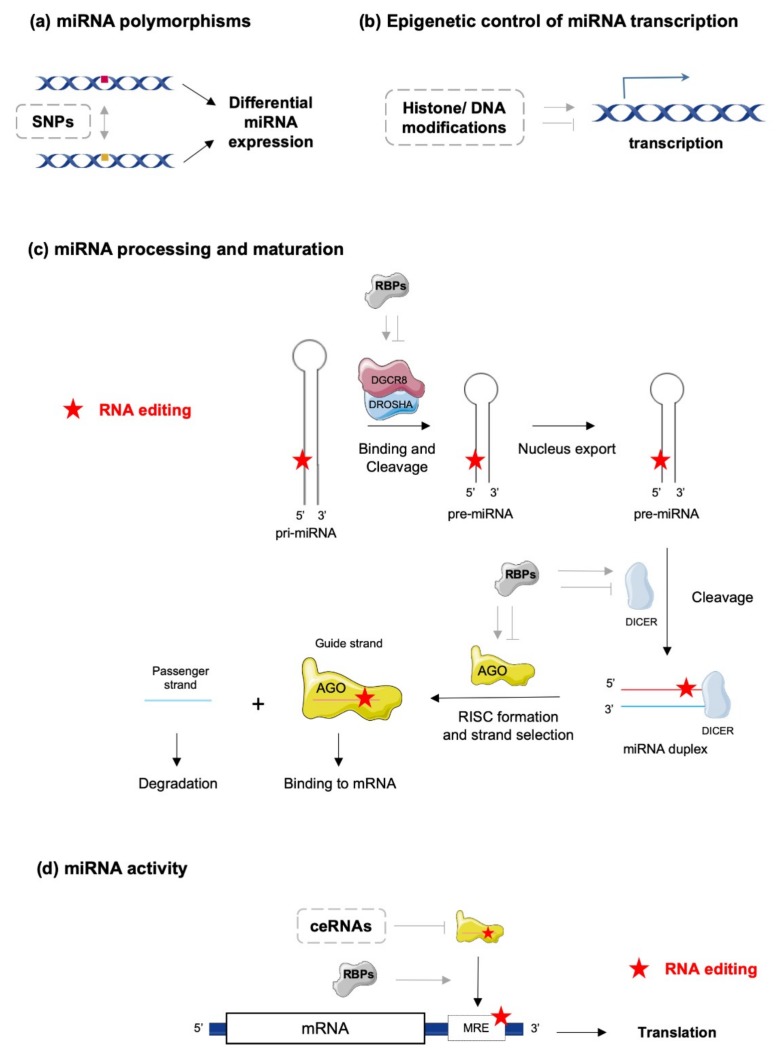
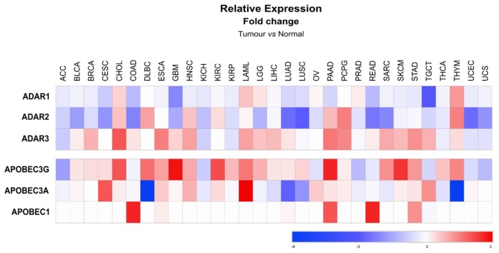
MicroRNAs (miRNAs) are small non-coding RNAs with the capability of modulating gene expression at the post-transcriptional level either by inhibiting messenger RNA (mRNA) translation or by promoting mRNA degradation. The outcome of a myriad of physiological processes and pathologies, including cancer, cardiovascular and metabolic diseases, relies highly on miRNAs. However, deciphering the precise roles of specific miRNAs in these pathophysiological contexts is challenging due to the high levels of complexity of their actions. Indeed, regulation of mRNA expression by miRNAs is frequently cell/organ specific; highly dependent on the stress and metabolic status of the organism; and often poorly correlated with miRNA expression levels. Such biological features of miRNAs suggest that various regulatory mechanisms control not only their expression, but also their activity and/or bioavailability. Several mechanisms have been described to modulate miRNA action, including genetic polymorphisms, methylation of miRNA promoters, asymmetric miRNA strand selection, interactions with RNA-binding proteins (RBPs) or other coding/non-coding RNAs. Moreover, nucleotide modifications (A-to-I or C-to-U) within the miRNA sequences at different stages of their maturation are also critical for their functionality. This regulatory mechanism called "RNA editing" involves specific enzymes of the adenosine/cytidine deaminase family, which trigger single nucleotide changes in primary miRNAs. These nucleotide modifications greatly influence a miRNA's stability, maturation and activity by changing its specificity towards target mRNAs. Understanding how editing events impact miRNA's ability to regulate stress responses in cells and organs, or the development of specific pathologies, e.g., metabolic diseases or cancer, should not only deepen our knowledge of molecular mechanisms underlying complex diseases, but can also facilitate the design of new therapeutic approaches based on miRNA targeting. Herein, we will discuss the current knowledge on miRNA editing and how this mechanism regulates miRNA biogenesis and activity.

Keywords: ADARs; APOBECs; miRNA editing; microRNA regulation.


Conflict of interest statement

The authors declare no conflict of interest.

Figures

Figure 1
Figure 1
Molecular mechanisms controlling microRNA (miRNA) biogenesis and activity. (a) Single nucleotide polymorphisms (SNPs) in genomic regions encoding miRNAs can impact their processing and maturation, resulting in different expression levels between variants. (b) Epigenetic modifications such as histone acetylation or DNA methylation can modulate transcription efficiencies of miRNAs. (c) RNA editing (red stars) and RNA-binding proteins (RBPs) can interfere with miRNA processing and maturation by affecting DGCR8/DROSHA activity, nuclear export of pre-miRNAs, cleavage of pre-miRNA by DICER and incorporation of mature miRNA in the RISC (AGO) complex. (d) Mature miRNA activity can be regulated by alternative mechanisms including (i) competition with RBPs for the same binding site on a target mRNA, (ii) decoying of the miRNA–RISC complex by ceRNAs and (iii) RNA editing of miRNA response elements (MREs) on target mRNAs. Stimulatory and inhibitory effects are represented by pointed and blunt grey arrows, respectively.
Figure 2
Figure 2
Expression patterns of ADAR and APOBEC editing enzymes. (a) Heat map showing mean relative expression (log2(transcripts per million (TPM) + 1)) of ADARs and APOBECs across different tissues. Expression levels of each gene were retrieved using GEPIA2 database (http://gepia2.cancer-pku.cn/#index), which include non-tumoral tissue samples from the Cancer Genome Atlas (TCGA) and normal tissue samples from the genotype-tissue expression (GTEx) projects. (b) Heat map showing mean relative expressions (log2 TPM + 1) of ADARs and APOBECs in subpopulations of immune cells, before or after activation. Expression levels of each gene were retrieved from DICE database (https://dice-database.org/). Both heat maps were designed using Morpheus (https://software.broadinstitute.org/morpheus).
Figure 3
Figure 3
Functional outcomes of RNA editing on miRNA biogenesis and activity. (a) ADAR-mediated miRNA editing can occur in pri- and pre-miRNA, both within and outside of seed sequences, thereby affecting their processing (pointed black arrows: stimulation of processing; blunt grey arrows: inhibition of processing) and final expression levels of the mature miRNA duplexes. Editing can also occur on mature miRNA duplexes seed sequences either modulating their activities or changing their specificities to different target mRNAs. (b) Activity of APOBEC enzymes interfere with miRNA functions through editing-dependent or independent mechanisms. Stimulatory and inhibitory effects are represented by pointed black and blunt grey arrows, respectively. APOBEC editing of single strand miRNAs associated with AGO can affect miRNA activity or change miRNA specificity to different target mRNAs (left side of the panel). On the other hand, APOBEC activity can also directly impact the RISC complex and perturb its assembly (right side of the panel). (c) APOBEC/ADAR-mediated miRNA editing can modulate miRNAs activity through different mechanisms. First, editing of the seed sequence on miRNAs can change their specificities for given mRNAs or ceRNAs (upper panels). Second, editing of the MRE on mRNA targets can either impede the recognition and binding of miRNAs (middle panels), or on the contrary, modify MREs, thereby allowing binding of miRNAs that were not recognizing the unedited MRE (lower panels).
Figure 4
Figure 4
Predicted human miRNAs potentially regulating ADARs and APOBECs expressions. (a) Predicted human miRNAs targeting the three ADAR isoforms. (b) Predicted human miRNAs targeting the three APOBEC isoforms. The lists of predicted miRNAs modulating ADARs or APOBECs were retrieved from the miRWalk 2.0 database. Predicted miRNAs were obtained using 12 different algorithms (i.e., miRWalk2.0, MicroT4, miRanda, miRBridge, miRDB, miRMap, miRNAMap, PICTAR2, PITA, RNA22, RNAhybrid and TargetScan). Only miRNAs predicted by at least five different algorithms were considered.
Figure 5
Figure 5
Expression patterns of ADAR and APOBEC editing enzymes in tumors. The heat map displays the mean fold change, following a log2 transformation, of ADAR and APOBEC gene expression in tumors from different tissues compared to their related non-tumoral tissues. Expression levels of each gene were retrieved using GEPIA2 database (http://gepia2.cancer-pku.cn/#index), which includes non-tumoral tissue and tumor samples from the Cancer Genome Atlas (TCGA). Fold changes were calculated using the ratio of tumor versus non-tumoral expression levels. Heat map was designed using Morpheus (https://software.broadinstitute.org/morpheus). Tumors are identified according with the TCGA nomenclature: ACC—adrenocortical carcinoma; BLCA—bladder urothelial carcinoma; BRCA—breast invasive carcinoma; CESC—cervical squamous cell carcinoma and endocervical adenocarcinoma; CHOL—cholangiocarcinoma; COAD—colon adenocarcinoma; DLBC—lymphoid neoplasm diffuse large B-cell lymphoma; ESCA—esophageal carcinoma; GBM—glioblastoma multiform; HNSC—head and neck squamous cell carcinoma; KICH—kidney chromophobe; KIRC—kidney renal clear cell carcinoma; KIRP—kidney renal papillary cell carcinoma; LAML – lymphoblastic acute myeloid leukemia; LGG—brain lower grade glioma; LIHC—liver hepatocellular carcinoma; LUAD—lung adenocarcinoma; LUSC—lung squamous cell carcinoma; MESO—mesothelioma; OV—ovarian serous cystadenocarcinoma; PAAD—pancreatic adenocarcinoma; PCPG—pheochromocytoma and paraganglioma; PRAD—prostate adenocarcinoma; READ—rectum adenocarcinoma; SARC—sarcoma; SKCM—skin cutaneous Melanoma; STAD—stomach adenocarcinoma; TGCT—testicular germ cell tumors; THCA—thyroid carcinoma; THYM—thymoma; UCEC—uterine corpus endometrial carcinoma; UCS—uterine carcinosarcoma; UVM—uveal melanoma.

References

    1. Vasudevan S., Tong Y., Steitz J.A. Switching from repression to activation: MicroRNAs can up-regulate translation. Science. 2007;318:1931–1934. doi: 10.1126/science.1149460. - DOI - PubMed
    1. Bartel D.P. MicroRNAs: Target recognition and regulatory functions. Cell. 2009;136:215–233. doi: 10.1016/j.cell.2009.01.002. - DOI - PMC - PubMed
    1. Nigita G., Acunzo M., Romano G., Veneziano D., Lagana A., Vitiello M., Wernicke D., Ferro A., Croce C.M. microRNA editing in seed region aligns with cellular changes in hypoxic conditions. Nucleic Acids Res. 2016;44:6298–6308. doi: 10.1093/nar/gkw532. - DOI - PMC - PubMed
    1. Turunen T.A., Roberts T.C., Laitinen P., Vaananen M.A., Korhonen P., Malm T., Yla-Herttuala S., Turunen M.P. Changes in nuclear and cytoplasmic microRNA distribution in response to hypoxic stress. Sci. Rep. 2019;9:10332. doi: 10.1038/s41598-019-46841-1. - DOI - PMC - PubMed
    1. Lee H.Y., Zhou K., Smith A.M., Noland C.L., Doudna J.A. Differential roles of human Dicer-binding proteins TRBP and PACT in small RNA processing. Nucleic Acids Res. 2013;41:6568–6576. doi: 10.1093/nar/gkt361. - DOI - PMC - PubMed

LinkOut - more resources