. 2019 Dec 11;8(12):637.
doi: 10.3390/antiox8120637.

Inhibitory Effect of β-Carotene on Helicobacter pylori-Induced TRAF Expression and Hyper-Proliferation in Gastric Epithelial Cells

Affiliations

Inhibitory Effect of β-Carotene on Helicobacter pylori-Induced TRAF Expression and Hyper-Proliferation in Gastric Epithelial Cells

Yongchae Park et al. Antioxidants (Basel). .

Abstract

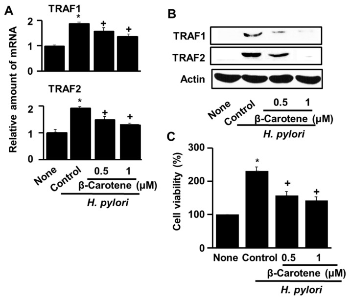
Helicobacter pylori infection causes the hyper-proliferation of gastric epithelial cells that leads to the development of gastric cancer. Overexpression of tumor necrosis factor receptor associated factor (TRAF) is shown in gastric cancer cells. The dietary antioxidant β-carotene has been shown to counter hyper-proliferation in H. pylori-infected gastric epithelial cells. The present study was carried out to examine the β-carotene mechanism of action. We first showed that H. pylori infection decreases cellular IBα levels while increasing cell viability, NADPH oxidase activity, reactive oxygen species production, nuclear factor kappa-light-chain-enhancer of activated B cells (NF-B) activation, and TRAF1 and TRAF2 gene expression, as well as protein-protein interaction in gastric epithelial AGS cells. We then demonstrated that pretreatment of cells with β-carotene significantly attenuates these effects. Our findings support the proposal that β-carotene has anti-cancer activity by reducing NADPH oxidase-mediated production of ROS, NF-B activation and NF-B-regulated TRAF1 and TRAF2 gene expression, and hyper-proliferation in AGS cells. We suggest that the consumption of β-carotene-enriched foods could decrease the incidence of H. pylori-associated gastric disorders.

Keywords: Helicobacter pylori; NF-B; hyper-proliferation; tumor necrosis factor receptor-associated factor; β-carotene.


Conflict of interest statement

The authors declare no conflict of interest.

Figures

Figure 1
Figure 1
H. pylori induces the expression of TRAF1 and TRAF2, IκBα degradation, NF-κB activation, and hyper-proliferation, but does not induce apoptosis in AGS cells. (A) The number of viable AGS cells measured using the trypan blue exclusion test at 0 h, 24 h, and 48 h following infection at a 1:20 and a 1:50 AGS cells-to-H. pylori ratio. *p < 0.05 vs. none (B) Flow cytometric analysis results of H. pylori-induced apoptosis in AGS cells following infection by H. pylori in a 1:20 and a 1:50 AGS cells to H. pylori ratio for 48 h (upper panel). A statistical graph of annexin V/PI staining is shown (lower panel). Apoptotic cells include the Annexin V+/PI− cells and the Annexin V+/PI+ cells. (C) The relative amounts of TRAF1 and TRAF2 mRNA in AGS cells measured by real time PCR at 0 h, 1 h, and 2 h following infection by H. pylori in a 1:50 AGS cells to H. pylori ratio (left panel). At 1 h-culture, mRNA expression of TRAF1 and TRAF2 was determined in uninfected cells (None”) or in H. pylori-infected cells in a 1:50 AGS cells to H. pylori ratio (“H. pylori”) (right panel). The mRNA levels were normalized to that of GAPDH. * p < 0.05 vs. 0 h or none. (D) Western blot analysis of the levels of TRAF1 and TRAF2 proteins in AGS cells following infection by H. pylori in a 1:50 ratio and for 2 h, 4 h and 8 h periods (upper panel). At 4 h of culture, protein expression of TRAF1 and TRAF2 was determined in uninfected cells (None”) or in H. pylori-infected cells in a 1:50 AGS cells to H. pylori ratio (“H. pylori”) (lower panel). Actin was used as a loading control. (E) Western blot analysis of the levels of IκBα at 0 h, 1, 2, and 4 h of AGS cells incubation with H. pylori in a 1:50 AGS cells to H. pylori ratio (upper panel). At 1 h-culture, the levels of IκBα was determined in uninfected cells (None”) or in H. pylori-infected cells in a 1:50 AGS cells to H. pylori ratio (“H. pylori”) (lower panel). Actin was used as a loading control. (F) EMSA analysis was used for determination of the amount of active nuclear NF-κB in AGS cells following infection by H. pylori in a 1:50 ratio and for 0.5 h and 1 h periods (left panel). At 1 h-culture, active nuclear NF-κB levels were determined in uninfected cells (None”) or in H. pylori-infected cells in a 1:50 AGS cells to H. pylori ratio (“H. pylori”) (right panel). “Free probe” corresponds to the [32P]-oligonucleotide probe used in the shift assay.
Figure 2
Figure 2
β-Carotene inhibits H. pylori-induced TRAF1 and TRAF2 gene expression and hyper-proliferation in AGS cells. The cells were pretreated with the indicated concentrations of β-carotene for 2 h, and then infected with H. pylori at a titer ratio of 1:50. The column “None” corresponds to uninfected, untreated cells, the column “Control” corresponds to untreated cells infected with H. pylori and the columns “0.5” and “1” correspond to cells incubated with 0.5 and 1.0 μM β-carotene, respectively prior to infection with H. pylori. (A) The relative amounts of TRAF1 and TRAF2 mRNA in AGS cells measured by real time PCR without H. pylori infection or with H. pylori infection at 1 h culture (at the ratio of AGS cells with H. pylori, 1:50). The mRNA levels were normalized to GAPDH mRNA. * p < 0.05 vs. none, + p < 0.05 vs. control. (B) Western blot analysis of TRAF1 and TRAF2 in AGS cells without H. pylori infection, or with H. pylori infection in the absence and presence of β-carotene at 4 h of culture. Actin was used as a loading control. (C) The number of viable AGS cells measured without H. pylori infection or with H. pylori infection in the absence and presence of β-carotene at 48 h of culture. The cell viability was measured using the MTT assay. * p < 0.05 vs. none, + p < 0.05 vs. control.
Figure 3
Figure 3
β-Carotene inhibits the effects of H. pylori infection of AGS cells on cellular ROS, NADPH oxidase, IκBα, and NF-κB. The cells were pretreated with the indicated concentrations of β-carotene for 2 h, and then infected with H. pylori at the titer ratio of 1:50 for 1 h. The column “None” corresponds to uninfected cells, the column “Control” corresponds to untreated cells infected with H. pylori, and the columns “0.5” and “1” correspond to cells incubated with 0.5 and 1 μM β-carotene, respectively, prior to infection with H. pylori. (A) ROS levels determined with the DCF assay. * p < 0.05 vs. none, + p < 0.05 vs. control. (B) NADPH oxidase activity determined with the Lucigenin assay. * p < 0.05 vs. none, + p < 0.05 vs. control. (C) IκBα levels determined by western blot analysis. Actin was used as the loading control. (D) NF-κB DNA-binding activity determined with the EMSA assay.
Figure 4
Figure 4
Transfection of AGS cells with an IκBα mutant gene inhibits H. pylori-induced expression of TRAF1 and TRAF2 and NF-κB activation. AGS cells that were transfected for 16 h with the control vector (“pcDNA”), or with the plasmid containing the IκBα mutant gene (“IκBα mt”), and then incubated with H. pylori at the titer ratio of 1:50 for 4 h (A), or 1 h (B). “H. pylori” corresponds to samples derived from infected cells, whereas “None” corresponds to samples derived from uninfected cells. (A) Western blot analysis of the levels of TRAF1 and TRAF2 with actin serving as the loading control. (B) The nuclear NF-κB DNA binding activity determined using the EMSA assay.
Figure 5
Figure 5
Transfection with the dominant negative mutant TRAF1 gene decreases the interaction of TRAF1 and TRAF2 and cell proliferation in H. pylori-infected AGS cells. The cells were transfected with control vector (“pcDNA”) or plasmid containing the dominant negative mutant TRAF1 gene (“TRAF1 mt”) for 16 h and then cultured with (“H. pylori”) or without (“None”) H. pylori at the titer ratio of 1:50 for 4 h (A) or 48 h (B). (A) Western blot analysis of the TRAF1- or TRAF2-antibody immunoprecipitate from whole-cell extracts of the cells transfected with control vector (“pcDNA”) or dominant negative mutant TRAF1 expression vector (“TRAF1 mt”), using TRAF1 and TRAF2 antibodies for visualization (upper panel). The cellular lysate (“Input”) served as the protein expression control (lower panel). (B) The percentage ratio of viable AGS cells transfected with control vector (“pcDNA”) or dominant negative mutant TRAF1 expression vector (“TRAF1 mt”) and in culture with vs. without H. pylori. The cell viability was measured using the MTT assay. * p < 0.05 vs. corresponding none.

References

    1. Tonkic A., Tonkic M., Lehours P., Mégraud F. Epidemiology and diagnosis of Helicobacter pylori infection. Helicobacter. 2012;17:1–8. doi: 10.1111/j.1523-5378.2012.00975.x. - DOI - PubMed
    1. Keilberg D., Ottemann K.M. How Helicobacter pylori senses, targets and interacts with the gastric epithelium. Environ. Microbiol. 2016;18:791–806. doi: 10.1111/1462-2920.13222. - DOI - PubMed
    1. Subhash V.V., Ho B. Inflammation and proliferation—A causal event of host response to Helicobacter pylori infection. Microbiology. 2015;161:1150–1160. doi: 10.1099/mic.0.000066. - DOI - PubMed
    1. Byun E., Lim J.W., Kim J.M., Kim H. α-Lipoic acid inhibits Helicobacter pylori-induced oncogene expression and hyperproliferation by suppressing the activation of NADPH oxidase in gastric epithelial cells. Med. Inflamm. 2014;2014:380830. doi: 10.1155/2014/380830. - DOI - PMC - PubMed
    1. Jang S.H., Lim J.W., Kim H. Beta-carotene inhibits Helicobacter pylori-induced expression of inducible nitric oxide synthase and cyclooxygenase-2 in human gastric epithelial AGS cells. J. Physiol. Pharmacol. 2009;7:131–137. - PubMed

LinkOut - more resources