. 2020 Feb 19;105(4):742-758.e6.
doi: 10.1016/j.neuron.2019.11.012. Epub 2019 Dec 10.

Integrative and Network-Specific Connectivity of the Basal Ganglia and Thalamus Defined in Individuals

Affiliations

Integrative and Network-Specific Connectivity of the Basal Ganglia and Thalamus Defined in Individuals

Deanna J Greene et al. Neuron. .

Abstract

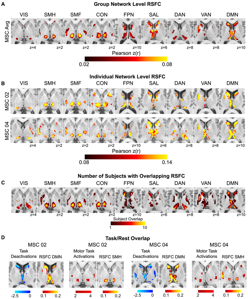
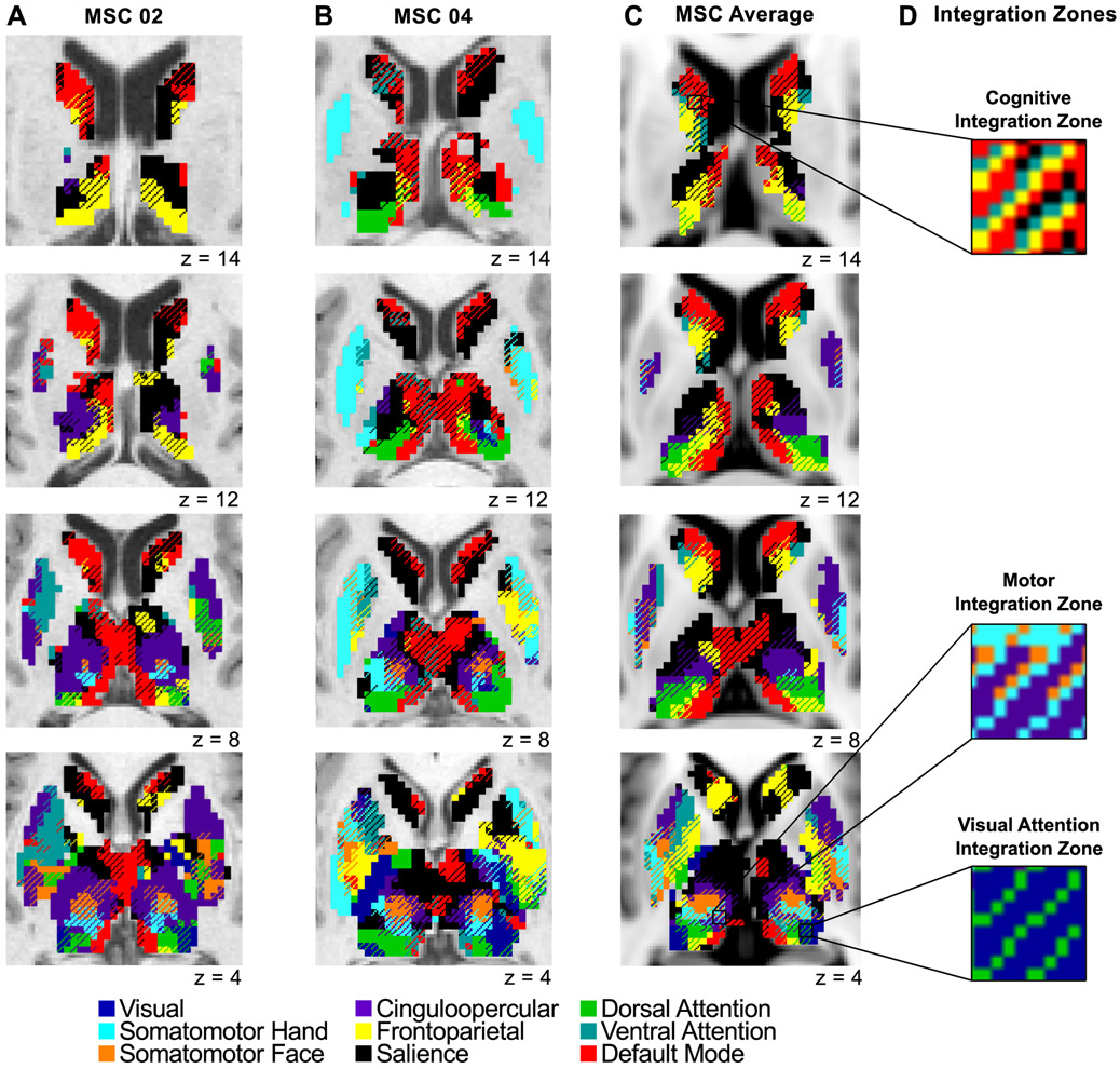
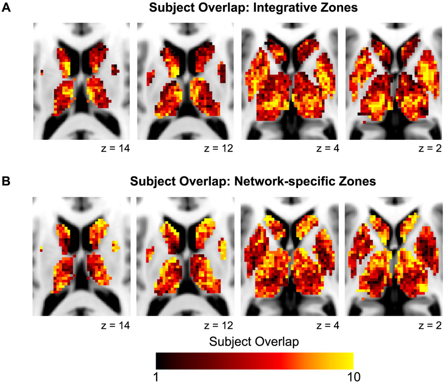
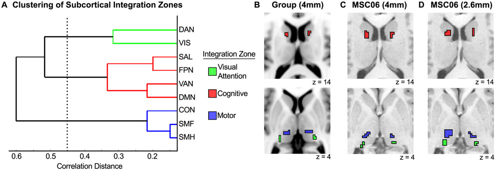
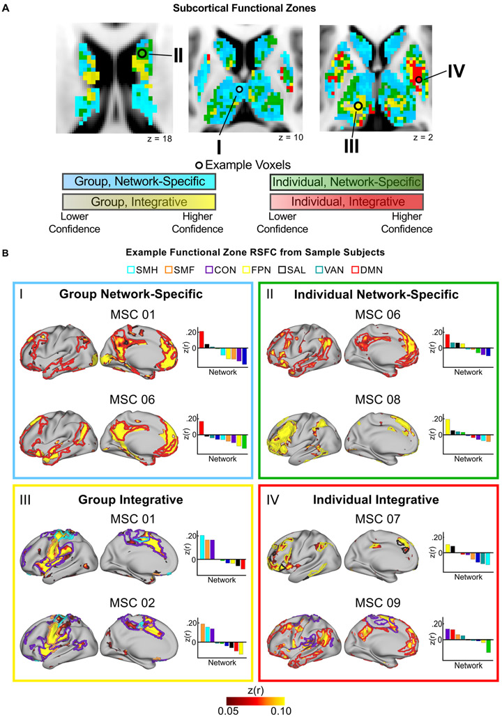
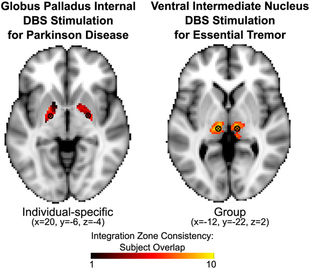
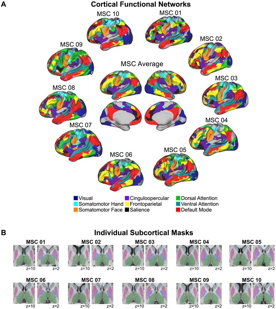
The basal ganglia, thalamus, and cerebral cortex form an interconnected network implicated in many neurological and psychiatric illnesses. A better understanding of cortico-subcortical circuits in individuals will aid in development of personalized treatments. Using precision functional mapping-individual-specific analysis of highly sampled human participants-we investigated individual-specific functional connectivity between subcortical structures and cortical functional networks. This approach revealed distinct subcortical zones of network specificity and multi-network integration. Integration zones were systematic, with convergence of cingulo-opercular control and somatomotor networks in the ventral intermediate thalamus (motor integration zones), dorsal attention and visual networks in the pulvinar, and default mode and multiple control networks in the caudate nucleus. The motor integration zones were present in every individual and correspond to consistently successful sites of deep brain stimulation (DBS; essential tremor). Individually variable subcortical zones correspond to DBS sites with less consistent treatment effects, highlighting the importance of PFM for neurosurgery, neurology, and psychiatry.

Keywords: basal ganglia; brain networks; deep brain stimulation; functional connectivity; precision functional mapping; resting state; subcortex; thalamus.


Conflict of interest statement

Declaration of Interests The authors declare no competing interests.

Figures

Figure 1.
Figure 1.. Framework for subcortical functional organization.
Subcortical RSFC was characterized along two principal axes, each with two levels (Group vs. Individual; Network specific vs. Integrative). “Group” refers to RSFC that is common across participants, whereas “Individual” refers to RSFC that is individually-specific. “Network specific” refers to regions of the subcortex with preferential RSFC to a single cortical network, whereas “Integrative” refers to regions of the subcortex with preferential RSFC to multiple cortical networks.
Figure 2.
Figure 2.. Subcortical RSFC is measurable at the individual level.
(A) Group-averaged subcortical RSFC for each cortical network. (B) Individual-level subcortical RSFC for each network from two representative MSC subjects, one male (MSC02) and one female (MSC04) (see Fig. S2 for all ten subjects). (C) Subject overlap showing the number of subjects with strong (top 10%) correlations with each network at that voxel. (D) Concordance between RSFC and task activations/deactivations within individuals. Task-evoked increases in BOLD activity during a motor task converge with peak RSFC in the somatomotor hand network. Task-evoked deactivations during a set of cognitive/perceptual tasks converge with peak RSFC in the default-mode network. Anatomical left is image left. VIS = visual; SMH = somatomotor hand; SMF = somatomotor face; CON = cingulo-opercular network; FPN = frontoparietal network; SAL = salience; DAN = dorsal attention network; VAN = ventral attention network; DMN = default-mode network.
Figure 3.
Figure 3.. Network-specific and integrative functional zones in the basal ganglia and thalamus.
Data displayed for (A) one male representative subject (MSC02), (B) one female representative subject (MSC04), and (C) the group average (all subjects shown in Fig. S7). Voxels with preferential RSFC to one network (network-specific) are represented by solid colors, and voxels functionally connected to multiple networks (integrative) are represented by cross-hatching. Anatomical left is image left. (D) Zooming in on several integration zones; three distinct clusters of integration zones (see Fig. 5): cognitive integration zones, motor integration zones, and visual attention integration zones.
Figure 4.
Figure 4.. Overlap across individuals of integrative and network-specific functional zones.
Higher values represent voxels with (A) integrative functional zones present in multiple subjects and (B) network-specific functional zones present in multiple subjects.
Figure 5.
Figure 5.. Three clusters of network integration are present in the subcortex.
(A) Hierarchical clustering revealed three clusters of network integration involving (1) dorsal attention and visual networks, (2) salience, frontoparietal, ventral attention, and default-mode networks, and (3) cingulo-opercular, somatomotor face, and somatomotor hand networks. (B) Most prominent locus of each cluster for the group average. (C) Most prominent locus of each cluster for an example individual (MSC06) with 4mm resolution data. (D) Most prominent locus of each cluster for the same individual (MSC06) with 2.6mm resolution data.
Figure 6.
Figure 6.. The subcortex contains four distinct functional zones: Group Network-Specific, Group Integrative, Individual Network-Specific, Individual Integrative.
(A) Anatomical distribution of each functional zone. Color gradation displays the confidence of zone assignment for each voxel as estimated by a jack-knifing procedure. (B) Typical examples of each type of functional zone using “example voxels” from panel A. Colored borders on the cortical surface represent the outline of the individual’s cortical networks that show strong RSFC with the example voxel. Bar graphs display RSFC correlations between the example voxel and each cortical network.
Figure 7.
Figure 7.. Overlap of integration zones and common sites of DBS.
Sites of DBS are shown with commonly targeted coordinates overlaid onto individual-specific (globus pallidus) and group (ventral intermediate thalamus) functional zones from the present study. Color gradation shows the consistency of the integration zones across subjects. The globus pallidus site, which has variable success rates, overlaps with an Individual Integrative zone. The ventral intermediate thalamus site, which has consistently high success rates, overlaps with a Group Integrative zone.
Figure 8.
Figure 8.. Functional cortical networks and subcortical voxels for each individual.
(A) Individually-defined functional networks (defined as in Gordon et al., 2017) and the group average functional networks are shown. Nine previously well-characterized functional networks were selected in order to investigate cortico-subcortical functional connectivity involving cortical networks that are described consistently using different methods and by multiple investigator groups (e.g., Damoiseaux et al., 2006; Gordon et al., 2016; Power et al., 2011; Yeo et al., 2011). Uncolored regions correspond to vertices that were not part of these nine networks according to the InfoMap network assignments. Note that including all 15 InfoMap networks (excluding unassigned and medial temporal vertices) did not change the results. (B) Subcortical masks from Freesurfer and manually edited using Freeview are shown for each individual. Light blue = caudate; pink = putamen, violet = pallidum; green = thalamus.

Comment in

References

    1. Albin RL, Young AB, and Penney JB (1989). The functional anatomy of basal ganglia disorders. Trends Neurosci 12, 366–375. - PubMed
    1. Alexander GE, and Crutcher MD (1990a). Basal ganglia-thalamocortical circuits: parallel substrates for motor,oculomotor,prefrontal and limbic functions. Prog Brain Res 85, 119–146. - PubMed
    1. Alexander GE, and Crutcher MD (1990b). Functional architecture of basal ganglia circuits: neural substrates of parallel processing. Trends Neurosci 13, 266–271. - PubMed
    1. Alexander GE, Delong MR, and Strick PL (1986). Parallel organization of functionally segregated circuits linking basal ganglia and cortex. Annu Rev Neurosci 9, 357–381. - PubMed
    1. Alhourani A, McDowell MM, Randazzo MJ, Wozny TA, Kondylis ED, Lipski WJ, Beck S, Karp JF, Ghuman AS, and Richardson RM (2015). Network effects of deep brain stimulation. J Neurophysiol 114, 2105–2117. - PMC - PubMed

Publication types