. 2020 Jan:138:304-317.
doi: 10.1016/j.yjmcc.2019.11.159. Epub 2019 Dec 10.

Defining decreased protein succinylation of failing human cardiac myofibrils in ischemic cardiomyopathy

Affiliations

Defining decreased protein succinylation of failing human cardiac myofibrils in ischemic cardiomyopathy

Hadi R Ali et al. J Mol Cell Cardiol. 2020 Jan.

Abstract

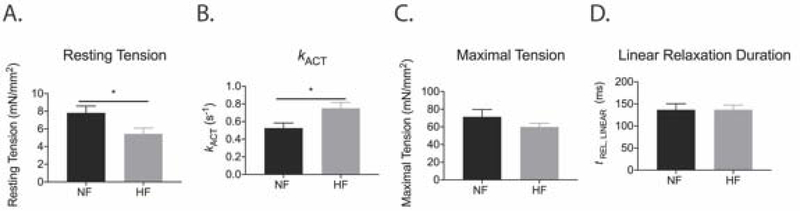
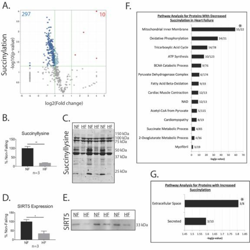
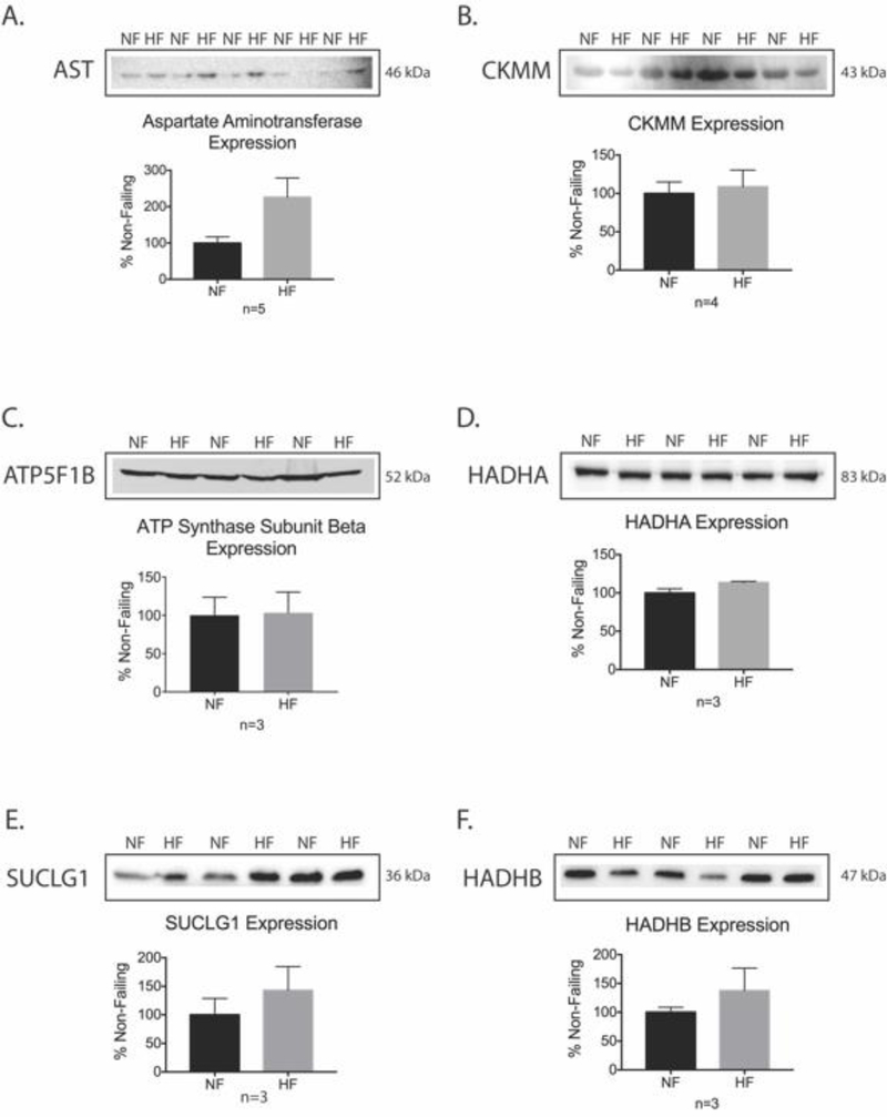
Succinylation is a post-translational modification of protein lysine residues with succinyl groups derived from succinyl CoA. Succinylation is considered a significant post-translational modification with the potential to impact protein function which is highly conserved across numerous species. The role of succinylation in the heart, especially in heart failure and myofibril mechanics, remains largely unexplored. Mechanical parameters were measured in myofibrils isolated from failing hearts of ischemic cardiomyopathy patients and non-failing donor controls. We employed mass spectrometry to quantify differential protein expression in myofibrils from failing ischemic cardiomyopathy hearts compared to non-failing hearts. In addition, we combined peptide enrichment by immunoprecipitation with liquid chromatography tandem mass spectrometry to quantitatively analyze succinylated lysine residues in these myofibrils. Several key parameters of sarcomeric mechanical interactions were altered in myofibrils isolated from failing ischemic cardiomyopathy hearts, including lower resting tension and a faster rate of activation. Of the 100 differentially expressed proteins, 46 showed increased expression in ischemic heart failure, while 54 demonstrated decreased expression in ischemic heart failure. Our quantitative succinylome analysis identified a total of 572 unique succinylated lysine sites located on 181 proteins, with 307 significantly changed succinylation events. We found that 297 succinyl-Lys demonstrated decreased succinylation on 104 proteins, while 10 residues demonstrated increased succinylation on 4 proteins. Investigating succinyl CoA generation, enzyme activity assays demonstrated that α-ketoglutarate dehydrogenase and succinate dehydrogenase activities were significantly decreased in ischemic heart failure. An activity assay for succinyl CoA synthetase demonstrated a significant increase in ischemic heart failure. Taken together, our findings support the hypothesis that succinyl CoA production is decreased and succinyl CoA turnover is increased in ischemic heart failure, potentially resulting in an overall decrease in the mitochondrial succinyl CoA pool, which may contribute to decreased myofibril protein succinylation in heart failure.

Keywords: Heart failure; Myofibrils; Post-translational modification; Proteomics; Sirtuins; Succinylation.


Conflict of interest statement

Declaration of Competing Interest The authors have no conflict of interest to declare.

Figures

Figure 1.
Figure 1.. Mechanical parameters of myofibrils isolated from ischemic HF patients are distinct from myofibrils isolated from NF hearts.
(A) Resting tension was decreased in ischemic HF myofibrils compared to NF myofibrils (p < 0.05). (B) Ischemic HF myofibrils had faster activation of contraction (kACT) than NF myofibrils (p < 0.05). (C and D) There were no differences in maximal tension or the duration of linear phase relaxation between ischemic HF and NF myofibrils. Statistical analyses of normal data for A-D were assessed by Student’s t-test with Welch’s correction [N = 8 NF patients (6–13 myofibril measured per patient), 9 ischemic HF patients (5–16 myofibrils measured per patient)]. See raw data in Supplemental Table S2.
Figure 2.
Figure 2.. Differential protein expression in ischemic failing and non-failing human cardiac myofibrils.
(A) LC-MS/MS total protein quantification for ischemic failing vs non-failing human cardiac myofibrils demonstrating the number of differentially expressed proteins in ischemic HF vs NF samples (n = 5, log2(fold change) ≥ 1.5 or ≤ −1.5, p < 0.05). (B) Western blot confirmation of increased periostin expression in ischemic HF samples using Student’s t-test (mean ± SEM, n = 4, p < 0.005). (C) Western blot confirmation of decreased IDH2 expression in ischemic HF samples (mean ± SEM, n = 5, p < 0.05). (D) Western blot confirmation of increased ACAT1 expression in ischemic HF samples (mean ± SEM, n = 3, p < 0.005). (E) Pathway analysis of proteins with increased expression in ischemic HF. (F) Pathway analysis of proteins with decreased expression in ischemic HF. (G) Comparison of differentially expressed proteins in human ischemic HF identified by LC-MS/MS with differentially expressed proteins and genes from previous human heart failure studies. *For E and F, the first number after each bar on the graph represents the number of proteins identified per pathway. The second number represents the fold enrichment for that pathway.
Figure 3.
Figure 3.. Myofibril and mitochondrial protein succinylation are altered in ischemic heart failure.
(A) Volcano plot showing the number of peptides with significant changes in succinylation for ischemic HF vs NF samples (n = 4, log2(fold change) ≥ 1.5 or ≤ −1.5, p < 0.05). (B) Densitometry for anti-succinyllysine western blot using Student’s t-test (mean ± SEM, n = 3, **p < 0.005). (C) Representative anti-succinyllysine western blot. (D) Densitometry for anti-SIRT5 western blot (mean ± SEM, n = 3, *p < 0.05). (E) Representative SIRT5 Western blot. (F) Pathway analysis of myofibril proteins with decreased succinylation in ischemic HF samples. (G) Pathway analysis of myofibril proteins with increased succinylation in ischemic HF samples. *For F and G, the first number after each bar on the graph represents the number of succinylated proteins identified per pathway. The second number represents the fold enrichment for that pathway.
Figure 4.
Figure 4.. Western blot validation of specific HPLC-MS/MS total protein results for proteins with decreased succinylation.
(A-F) Representative western blot images and corresponding densitometry quantification using Student’s t-test for aspartate aminotransferase, creatine kinase M-type (CKMM), ATP synthase subunit beta (ATP5F1B), trifunctional enzyme subunit alpha (HADHA), succinyl CoA ligase [GDP forming] subunit alpha (SUCLG1), trifunctional enzyme subunit beta (HADHB) (mean ± SEM, *p < 0.05)
Figure 5.
Figure 5.. Proteomic map of decreased succinylation in ischemic HF samples.
Map includes proteins with succinylation changes ≤ −1.5, p < 0.05, which demonstrate decreased succinylation independent of changes in protein expression.
Figure 6.
Figure 6.. Enzyme activity assays demonstrate decreased TCA cycle function, decreased succinyl-CoA production, and increased succinyl CoA turnover in the ischemic failing heart
(A) SDH activity was significantly decreased, but expression of the SDHA subunit was unchanged in failing ICM hearts compared to NF hearts (mean ± SEM, n = 3, *p < 0.05). (B) SCS activity was significantly increased, but expression of the SUCLG1 subunit was unchanged in failing ICM hearts compared to NF hearts (mean ± SEM, n = 3, *p < 0.05). (C) α-KGDH activity was significantly decreased, but expression of the DLST subunit was unchanged in failing ICM hearts (mean ± SEM, n = 3, *p < 0.001). Statistical significance for A-C was assessed using Student’s t-test. (D) LC-MS/MS total protein quantification demonstrated no change in expression for the SDHA subunit of SDH, the SUCLG1 subunit of SCS, and the DLST subunit of α-KGDH. (E) LC-MS/MS protein succinylation quantification revealed that succinylation of SDH, SCS, and α-KGDH subunits were decreased.

References

    1. Klabunde RE. Cardiovascular physiology concepts. 2nd ed Philadelphis, PA: Lippincott Williams &Wilkins/Wolters Kluwer; 2011. 2011.
    1. Mozaffarian D, Benjamin EJ, Go AS, Arnett DK, Blaha MJ, Cushman M, et al. Heart Disease and Stroke Statistics—2015 Update: A Report From the American Heart Association. Circulation. 2015;131(4):e29–e322. - PubMed
    1. Benjamin EJ, Blaha MJ, Chiuve SE, Cushman M, Das SR, Deo R, et al. Heart Disease and Stroke Statistics-2017 Update: A Report From the American Heart Association. Circulation. 2017;135(10):e146–e603. - PMC - PubMed
    1. Mamas MA, Sperrin M, Watson MC, Coutts A, Wilde K, Burton C, et al. Do patients have worse outcomes in heart failure than in cancer? A primary care-based cohort study with 10-year follow-up in Scotland. European journal of heart failure. 2017;19(9):1095–104. - PubMed
    1. Taylor CJ, Ryan R, Nichols L, Gale N, Hobbs FR, Marshall T. Survival following a diagnosis of heart failure in primary care. Family practice. 2017;34(2):161–8. - PMC - PubMed

Publication types