. 2020 Feb 18;86(5):e02749-19.
doi: 10.1128/AEM.02749-19. Print 2020 Feb 18.

Melanin Produced by the Fast-Growing Marine Bacterium Vibrio natriegens through Heterologous Biosynthesis: Characterization and Application

Affiliations

Melanin Produced by the Fast-Growing Marine Bacterium Vibrio natriegens through Heterologous Biosynthesis: Characterization and Application

Zheng Wang et al. Appl Environ Microbiol. .

Abstract

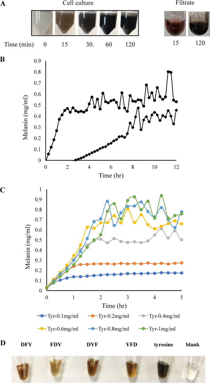
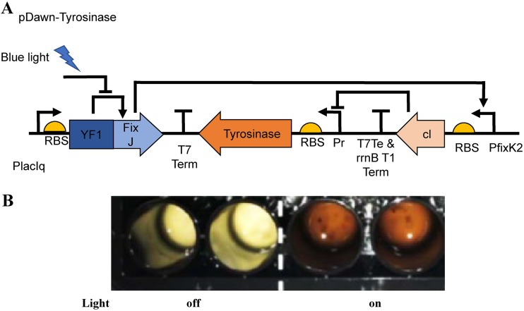
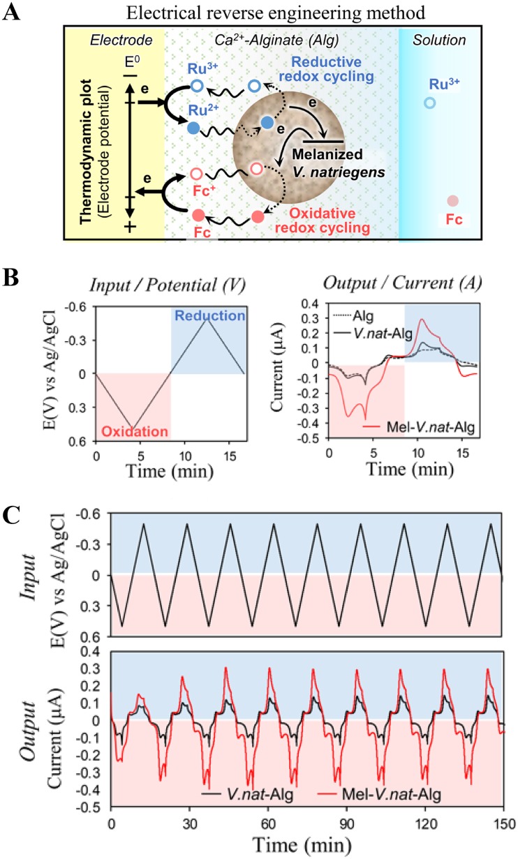
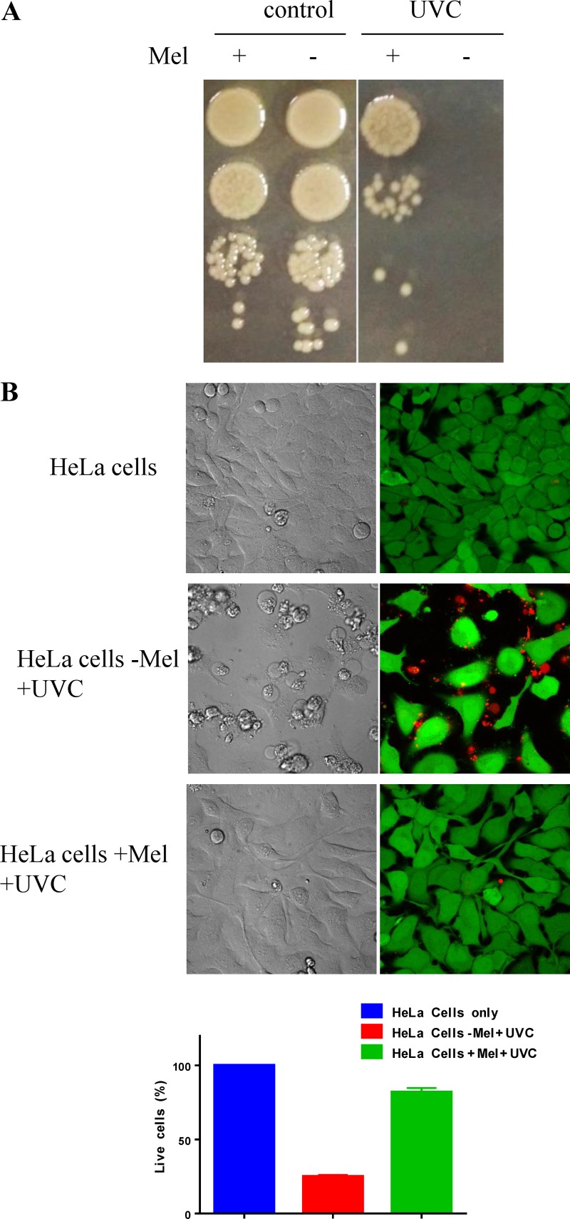
Melanin is a pigment produced by organisms throughout all domains of life. Due to its unique physicochemical properties, biocompatibility, and biostability, there has been an increasing interest in the use of melanin for broad applications. In the vast majority of studies, melanin has been either chemically synthesized or isolated from animals, which has restricted its use to small-scale applications. Using bacteria as biocatalysts is a promising and economical alternative for the large-scale production of biomaterials. In this study, we engineered the marine bacterium Vibrio natriegens, one of the fastest-growing organisms, to synthesize melanin by expressing a heterologous tyrosinase gene and demonstrated that melanin production was much faster than in previously reported heterologous systems. The melanin of V. natriegens was characterized as a polymer derived from dihydroxyindole-2-carboxylic acid (DHICA) and, similarly to synthetic melanin, exhibited several characteristic and useful features. Electron microscopy analysis demonstrated that melanin produced from V. natriegens formed nanoparticles that were assembled as "melanin ghost" structures, and the photoprotective properties of these particles were validated by their protection of cells from UV irradiation. Using a novel electrochemical reverse engineering method, we observed that melanization conferred redox activity to V. natriegens Moreover, melanized bacteria were able to quickly adsorb the organic compound trinitrotoluene (TNT). Overall, the genetic tractability, rapid division time, and ease of culture provide a set of attractive properties that compare favorably to current E. coli production strains and warrant the further development of this chassis as a microbial factory for natural product biosynthesis.IMPORTANCE Melanins are macromolecules that are ubiquitous in nature and impart a large variety of biological functions, including structure, coloration, radiation resistance, free radical scavenging, and thermoregulation. Currently, in the majority of investigations, melanins are either chemically synthesized or extracted from animals, which presents significant challenges for large-scale production. Bacteria have been used as biocatalysts to synthesize a variety of biomaterials due to their fast growth and amenability to genetic engineering using synthetic biology tools. In this study, we engineered the extremely fast-growing bacterium V. natriegens to synthesize melanin nanoparticles by expressing a heterologous tyrosinase gene with inducible promoters. Characterization of the melanin produced from V. natriegens-produced tyrosinase revealed that it exhibited physical and chemical properties similar to those of natural and chemically synthesized melanins, including nanoparticle structure, protection against UV damage, and adsorption of toxic compounds. We anticipate that producing and controlling melanin structures at the nanoscale in this bacterial system with synthetic biology tools will enable the design and rapid production of novel biomaterials for multiple applications.

Keywords: Vibrio natriegens; biomanufacturing; fast growing; melanin; melanin biosynthesis; nanoparticle; synthetic biology.


Figures

FIG 1
FIG 1
Expression of tyrosinase gene and melanin production in V. natriegens through IPTG induction. (A) Left, melanin production in M9 medium supplemented with 40 μg/ml CuSO4 and 0.4 mg/ml l-tyrosine after tyrosinase was induced in rich medium; right, melanin production from the filtered supernatant supplemented with CuSO4 and l-tyrosine after the induced cells were incubated in M9 medium for 15 min. (B) Kinetics of melanin production in M9 (•) and LBv2 (♦) media supplemented with 40 μg/ml CuSO4 and 0.4 mg/ml l-tyrosine after tyrosinase was induced in rich medium. (C) Effect of l-tyrosine concentration (in milligrams per milliliter) on melanin production in M9 medium. Graphs in panels B and C represent the averages of the results from five independent experiments. (D) Syntheses of melanin variants with tripeptide precursors. IPTG-induced V. natriegens/pJV-Tyr1 cells were transferred into M9 medium supplemented with CuSO4 and 5 mM tripeptides and incubated at 37°C for 1 h.
FIG 2
FIG 2
Induction of tyrosinase gene expression and melanin production in V. natriegens in an optogenetic system. (A) pDawn plasmid for light-activated gene expression in V. natriegens. YF1/FixJ drives gene expression from the pfixK2 promoter and is repressed by blue light. Insertion of the λ phage repressor cI and the λ promoter pR makes expression of the tyrosinase gene light activated. (B) Melanin production in the light plate array. The engineered bacterial cells were incubated in each well for 48 h. Left, duplicate wells with light off; right, duplicate wells with light on. RBS, ribosome-binding site.
FIG 3
FIG 3
Extracellular melanin particles and bacterial melanin ghosts. (A) a, TEM image of melanin nanoparticles in the supernatant of V. natriegens cell culture after l-tyrosine addition in the bacterial culture at 30 min. b, hydrodynamic sizes of melanin nanoparticles measured by DLS. Melanin nanoparticles were formed after the addition of l-tyrosine in the presence of 250 mM NaCl. The measured average sizes were 16 ± 0.6 nm with l-tyrosine addition (similar to melanin nanoparticle measurement by TEM) and 97 ± 39 nm for the control without l-tyrosine addition that were attributed to the by-products of cell culture extract. c, melanin nanoparticles formed after l-tyrosine addition at 12 h with different concentrations of salt. The peaks showed at 35 ± 3.4 nm (250 mM NaCl, blue), 73 ± 5.3 nm (350 mM NaCl, orange), and 86 ± 9.6 nm (550 mM NaCl, gray). (B) SEM and TEM images of melanin ghosts and melanized bacterial cells.
FIG 4
FIG 4
Characterization of melanin produced from V. natriegens. (A) FTIR spectra of bacterial (Bac) melanin and synthetic (Syn) melanin. Peaks in common and their assignments are 1,625 cm−1, C=C vibration in aromatic (indole) ring; 1,722 cm−1, C=O carbonyl group vibration from carboxylic acid in DHICA; triple peaks at ∼2,900 cm−1, CH2 alkyl vibration; and broad peak centered at 3,415 cm−1, O-H hydroxyl vibration. a.u., arbitrary units. (B) EPR spectra of melanin (Mel) powders, dried melanized and nonmelanized V. natriegens (Vnat) cells, and melanin ghosts. (C) EPR changes of melanin ghosts and dried melanized cells responding to illumination. arb., arbitrary.
FIG 5
FIG 5
Redox activity of melanized V. natriegens (V.nat) revealed by reverse electrical engineering method. (A) Schematic shows that melanin can donate/accept electrons to/from mediators by oxidative/reductive redox cycling process (details are described in the supplemental material). (B) The imposed input potential (i.e., voltage) and observed output current response associated with Ru3+ and Fc mediators. (C) Long-term cyclic experiments test for the reversibility of redox-activity; steady amplifications are signatures of reversible and repeated oxidation and reduction of melanin. E(V), potential in volts.
FIG 6
FIG 6
Photoprotective properties of biosynthesized melanin. (A) Survival of the nonmelanized and melanized V. natriegens cell cultures irradiated with 450 mJ/cm2 UVC. (B) Survival of HeLa cells suspended in the supernatants of the nonmelanized (−Mel) and the melanized (+Mel) V. natriegens and irradiated with 4 mJ/cm2 UVC. The control includes HeLa cells only without irradiation. Live/dead staining is shown as green for live cells and red for dead cells.
FIG 7
FIG 7
(A and B) Time-dependent (A) and pH-dependent (B) adsorption of TNT by ∼5 × 108 nonmelanized (light gray) and melanized (dark gray) V. natriegens cells.

References

    1. d'Ischia M, Wakamatsu K, Cicoira F, Di Mauro E, Garcia-Borron JC, Commo S, Galván I, Ghanem G, Kenzo K, Meredith P, Pezzella A, Santato C, Sarna T, Simon JD, Zecca L, Zucca FA, Napolitano A, Ito S. 2015. Melanins and melanogenesis: from pigment cells to human health and technological applications. Pigment Cell Melanoma Res 28:520–544. doi:10.1111/pcmr.12393. - DOI - PubMed
    1. Kim YJ, Khetan A, Wu W, Chun SE, Viswanathan V, Whitacre JF, Bettinger CJ. 2016. Evidence of porphyrin-like structures in natural melanin pigments using electrochemical fingerprinting. Adv Mater 28:3173–3180. doi:10.1002/adma.201504650. - DOI - PubMed
    1. Schweitzer AD, Howell RC, Jiang Z, Bryan RA, Gerfen G, Chen CC, Mah D, Cahill S, Casadevall A, Dadachova E. 2009. Physico-chemical evaluation of rationally designed melanins as novel nature-inspired radioprotectors. PLoS One 4:e7229. doi:10.1371/journal.pone.0007229. - DOI - PMC - PubMed
    1. Patel RP, Okun MR, Edelstein LM, Cariglia N. 1974. Peroxidatic conversion of tyrosine to dopachrome. J Invest Dermatol 63:374–377. doi:10.1111/1523-1747.ep12680867. - DOI - PubMed
    1. Liu Y, Simon JD. 2003. Isolation and biophysical studies of natural eumelanins: applications of imaging technologies and ultrafast spectroscopy. Pigment Cell Res 16:606–618. doi:10.1046/j.1600-0749.2003.00098.x. - DOI - PubMed

Publication types

MeSH terms

Supplementary concepts

LinkOut - more resources