. 2019 Dec 13;10(12):951.
doi: 10.1038/s41419-019-2185-x.

NBS1 interacts with HP1 to ensure genome integrity

Affiliations

NBS1 interacts with HP1 to ensure genome integrity

Giuseppe Bosso et al. Cell Death Dis. .

Abstract

Heterochromatin Protein 1 (HP1) and the Mre11-Rad50-Nbs1 (MRN) complex are conserved factors that play crucial role in genome stability and integrity. Despite their involvement in overlapping cellular functions, ranging from chromatin organization, telomere maintenance to DNA replication and repair, a tight functional relationship between HP1 and the MRN complex has never been elucidated. Here we show that the Drosophila HP1a protein binds to the MRN complex through its chromoshadow domain (CSD). In addition, loss of any of the MRN members reduces HP1a levels indicating that the MRN complex acts as regulator of HP1a stability. Moreover, overexpression of HP1a in nbs (but not in rad50 or mre11) mutant cells drastically reduces DNA damage associated with the loss of Nbs suggesting that HP1a and Nbs work in concert to maintain chromosome integrity in flies. We have also found that human HP1α and NBS1 interact with each other and that, similarly to Drosophila, siRNA-mediated inhibition of NBS1 reduces HP1α levels in human cultured cells. Surprisingly, fibroblasts from Nijmegen Breakage Syndrome (NBS) patients, carrying the 657del5 hypomorphic mutation in NBS1 and expressing the p26 and p70 NBS1 fragments, accumulate HP1α indicating that, differently from NBS1 knockout cells, the presence of truncated NBS1 extends HP1α turnover and/or promotes its stability. Remarkably, an siRNA-mediated reduction of HP1α in NBS fibroblasts decreases the hypersensitivity to irradiation, a characteristic of the NBS syndrome. Overall, our data provide an unanticipated evidence of a close interaction between HP1 and NBS1 that is essential for genome stability and point up HP1α as a potential target to counteract chromosome instability in NBS patient cells.


Conflict of interest statement

The authors declare that they have no conflict of interest.

Figures

Fig. 1
Fig. 1. Drosophila HP1a physically interacts with the MRN complex.
a Co-immunoprecipitation assay from HP1a-FLAG-overexpressing S2 cell extracts showing that HP1a precipitates endogenous Rad50, Mre11, and Nbs proteins (Input, 10% of total extract). The asterisk indicates aspecific bands. b, c Pulldown assays from Nbs-HA expressing Drosophila S2 cells with GST-tagged-full length HP1a (HP1a WT) and b GST-HP1aCSD and GST-HP1a∆CSD or c GST-HP1aW200A and GST-HP1I191E mutant proteins. Note that while Nbs has been revealed with a commercial anti-HA antibody, Rad50 and Mre11 have been detected with anti-Rad50- and anti-Mre11-specific antibodies generated in our laboratory. Ponceau staining shows the amount of each GST-tagged HP1a protein used in this assay. See text and “Materials and methods” for further details.
Fig. 2
Fig. 2. Loss of the Drosophila MRN complex affects HP1a levels.
a WB analysis from Oregon R wild-type and nbs1, rad50∆5.1, and mre11Δ35K.1 mutant larval brains showing that HP1a levels are reduced when any of the MRN complex member is depleted. The anti-Giotto antibody has been used as a loading control. The asterisks (*) indicate aspecific bands. Note the absence of Nbs, Rad50, and Mre11 bands in the corresponding nbs1, rad50∆5.1, and mre11Δ35K.1 loss-of-function mutants that also reveals the specificity of the our anti-Nbs, anti-Mre11 (this study), and anti-Rad50 antibodies. b Quantification analysis of HP1a reduction from at least four independent WBs. (t Student’s Test; *p < 0.05; **p < 0.01). c Localization of HP1a in nbs1, rad50∆5.1, and mre11Δ35K.1 mutant brain metaphases. Bar: 10 μm d Quantification of anti-HP1a immunolocalization (t Student’s Test; *p < 0.01. Bars indicate ±S.D.
Fig. 3
Fig. 3. HP1a does not affect the expression of the MRN complex.
a WB from Su(var)2-5/HP1a mutant extracts and from control (OR) or HP1a-expressing Su(var)2-5-RFP protein extracts, revealed with anti- MRN complex and anti-HP1 antibodies. Anti-actin and anti-Giotto have been used as loading controls. The asterisks (*) indicate aspecific bands. b Quantification of chromosome break frequency in MRN mutants expressing HP1a-RFP (at Student’s Test; p < 0.01). STAs = Single Telomere Attachments; DTAs = Double Telomere Attachments.
Fig. 4
Fig. 4. NBS1 interacts with HP1α following DNA damage.
a WB analysis of cellular extracts from untreated and irradiated MRC5 fibroblasts after immunoprecipitation (IP) with either anti-HP1α or anti-NBS1 antibodies. MRC5 cells were irradiated with 5 Gy of X-rays and harvested after 0.5 h. One milligram of total protein extracts were immunoprecipitated and 10 μg of total protein lysate (1%) were loaded as inputs. Membranes were probed with anti-HP1α or anti-NBS1 antibodies. Note that HP1α cannot be revealed in the IP of NBS1 since its molecular weight of ~22 kDa is the same as the light chain of immunoglobulins. Total IgG levels and vinculin were used as loading control for immunoprecipitates and input, respectively. b Top: Structural complex between HP1α (sea green) and the NBS1 peptide containing the PGPSL sequence (coral) that allows the recognition of HP1α. Down: Structural complex between HP1α (sky blue) and the NBS1 peptide containing the PGPSL sequence (orange) in which the S343 residue is phosphorylated. c Electrostatic potential surface representation of the structural complex HP1α–NBS1 peptide in which the S343 residue is either unphosphorylated (top) or phosphorylated (down) (red, −2 kT/e; white, 0.0 kT/e; blue, +2 kT/e). d WB from control MRC5 cells and NBS1 cells carrying the S343A point mutation, immunoprecipitated with anti-HP1α antibody. Ten micrograms of total protein lysate (1%) were loaded as input. Membranes were blotted with anti-NBS1; total IgG levels and vinculin were used as loading controls for immunoprecipitates and input, respectively.
Fig. 5
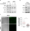
Fig. 5. NBS1 regulates the stability of HP1α.
a WB from both untreated and irradiated mocked and siNBS1– MRC5 cell extracts. Cells were irradiated with 5 Gy of X-rays and harvested after 0.5 h. Ten micrograms of total protein lysates were analyzed by WB. Membranes were probed with anti-HP1α and anti-NBS1 antibodies; vinculin and β-actin were used as loading controls. b WB from extracts of lymphoblastoid cells (LCLs) established from an healthy donor (Ctrl) and from a patient carrying two germline mutations in the RAD50 gene (RAD50−/−). Membranes were probed with anti-HP1α, anti-NBS1, and anti-RAD50 antibodies; β-actin was used as loading control. c Left: representative images of the immunofluorescence analysis of HP1α (Alexa Fluor 480, green fluorophore green) protein levels in mocked and NBS1-silecenced MRC5 cells. Bar: 20 μm. Right: Distribution of HP1α corrected nuclear fluorescence intensity in mocked and NBS1-silecenced MRC5 cells. The horizontal lanes indicate the mean values derived from the analysis of 100 cells/experimental point in three independent experiments ±S.D. (t Student’s Test; ***p < 0.001).
Fig. 6
Fig. 6. HP1α accumulates in NBS cells.
a Left: representative images of the immunofluorescence analysis of HP1 levels (green) in MRC5 and NBS cells. Bar: 20 μm. Right: Distribution of HP1α corrected nuclear fluorescence intensity in MRC5 and NBS1 cells. The horizontal lanes indicate the mean values derived from the analysis of 100 cells/experimental point in three independent experiments ±S.D. (t Student’s Test; ***p < 0.001). b WB analysis of HP1α levels in fibroblast and lymphoblastoid cells (LCLs). The levels of HP1α and NBS1 in NBS cells were compared to their corresponding controls (i.e., MRC5 for NBS1 fibroblasts; Ctrl for NBS1–1 and NBS1–2 lymphoblastoid cells); β-actin was used as the loading control.
Fig. 7
Fig. 7. NBS1 fragments arising from the 657del founder mutation in NBS1 patients increase HP1α levels.
a WB analysis from total protein lysate derived from NBS cells, immunoprecipitated with either anti-HP1α or with two different anti-NBS1 antibodies directed against the N- and the C-terminus of NBS1. Membranes were probed with the anti-HP1α or the two anti-NBS1 antibodies. The corresponding inputs (1%), probed with anti-HP1α and anti-vinculin antibodies, are shown below. b One milligram of whole-protein lysate obtained from NBS1 cells were incubated in the absence (−) or presence (+) of 10 U alkaline phosphatase (AP). The untreated and dephosphorylated samples were immunoprecipitated with anti-HP1α antibody and membranes were probed with anti-NBS1 antibody directed against the C-terminus of NBS1. Total IgG levels and vinculin were used as loading controls for immunoprecipitates and input, respectively.
Fig. 8
Fig. 8. HP1α inhibition reduces IR-induced DSBs in NBS cells.
Analysis of DSB repair kinetics of γH2AX and 53BP1 in mock- and HP1α-silenced MRC5 and NBS cells, irradiated with 1 Gy of X-rays and harvested after 0.5, 3, 6, and 24 h. Graphs express the mean number of either γH2AX or 53BP1 foci/cell derived from the analysis of 100 cells/experimental point, in three independent experiments ±S.D. (t Student’s Test; *p < 0.05, **p < 0.01, ***p < 0.001).

Similar articles

Cited by

References

    1. Maison C, Almouzni G. HP1 and the dynamics of heterochromatin maintenance. Nat. Rev. Mol. Cell Biol. 2004;5:296–304. doi: 10.1038/nrm1355. - DOI - PubMed
    1. Mendez DL, et al. The HP1a disordered C terminus and chromo shadow domain cooperate to select target peptide partners. Chembiochem. 2011;12:1084–1096. doi: 10.1002/cbic.201000598. - DOI - PMC - PubMed
    1. Liu Y, et al. Peptide recognition by heterochromatin protein 1 (HP1) chromoshadow domains revisited: Plasticity in the pseudosymmetric histone binding site of human HP1. J. Biol. Chem. 2017;292:5655–5664. doi: 10.1074/jbc.M116.768374. - DOI - PMC - PubMed
    1. Badugu R, Yoo Y, Singh PB, Kellum R. Mutations in the heterochromatin protein 1 (HP1) hinge domain affect HP1 protein interactions and chromosomal distribution. Chromosoma. 2005;113:370–384. doi: 10.1007/s00412-004-0324-2. - DOI - PubMed
    1. Fanti L, Pimpinelli S. HP1: a functionally multifaceted protein. Curr. Opin. Genet. Dev. 2008;18:169–174. doi: 10.1016/j.gde.2008.01.009. - DOI - PubMed

Publication types

MeSH terms

LinkOut - more resources