. 2019 Dec 13;9(1):19130.
doi: 10.1038/s41598-019-55240-5.

Fragment-based screening identifies molecules targeting the substrate-binding ankyrin repeat domains of tankyrase

Affiliations

Fragment-based screening identifies molecules targeting the substrate-binding ankyrin repeat domains of tankyrase

Katie Pollock et al. Sci Rep. .

Abstract

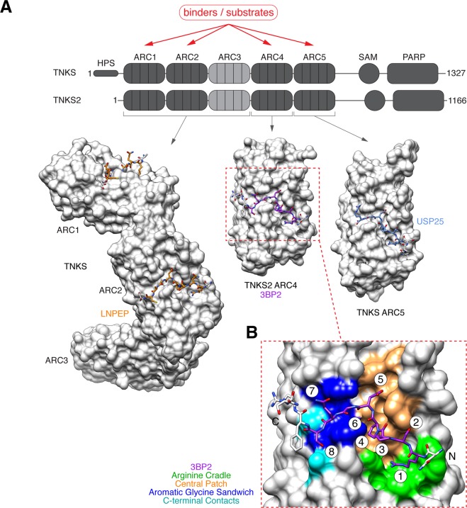
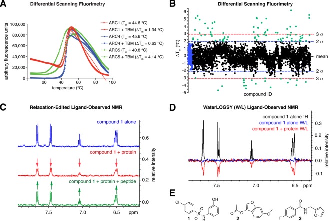
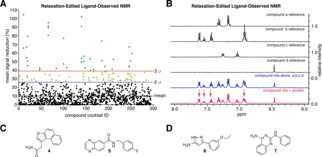
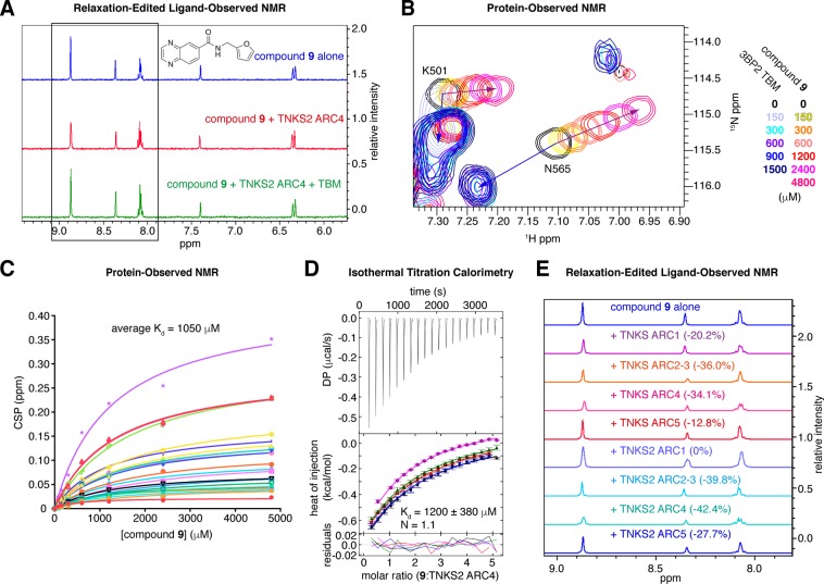
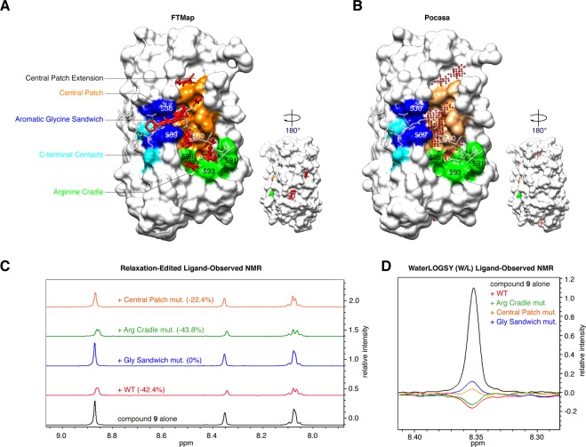
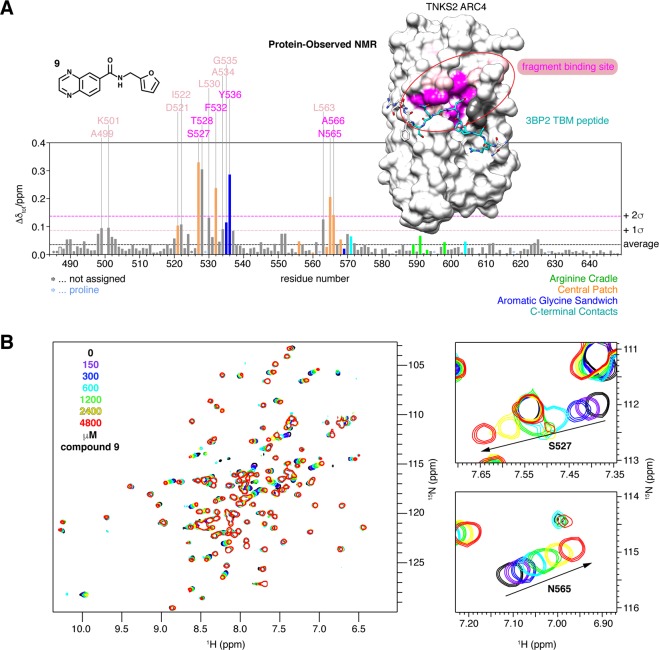
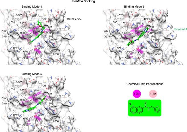
The PARP enzyme and scaffolding protein tankyrase (TNKS, TNKS2) uses its ankyrin repeat clusters (ARCs) to bind a wide range of proteins and thereby controls diverse cellular functions. A number of these are implicated in cancer-relevant processes, including Wnt/β-catenin signalling, Hippo signalling and telomere maintenance. The ARCs recognise a conserved tankyrase-binding peptide motif (TBM). All currently available tankyrase inhibitors target the catalytic domain and inhibit tankyrase's poly(ADP-ribosyl)ation function. However, there is emerging evidence that catalysis-independent "scaffolding" mechanisms contribute to tankyrase function. Here we report a fragment-based screening programme against tankyrase ARC domains, using a combination of biophysical assays, including differential scanning fluorimetry (DSF) and nuclear magnetic resonance (NMR) spectroscopy. We identify fragment molecules that will serve as starting points for the development of tankyrase substrate binding antagonists. Such compounds will enable probing the scaffolding functions of tankyrase, and may, in the future, provide potential alternative therapeutic approaches to inhibiting tankyrase activity in cancer and other conditions.


Conflict of interest statement

The authors declare no competing interests.

Figures

Figure 1
Figure 1
(A) Domain organisation of human tankyrase enzymes. Two tankyrase paralogues (TNKS, TNKS2) share an overall sequence identity of 82% (83% across ARCs, 74% across SAM domains, 94% across PARP domains). The ARCs comprise the substrate/protein recognition domain. Several examples of crystal structures of human tankyrase ARCs bound to tankyrase-binding motif (TBM) peptides are shown: TNKS ARC1-3 bound to TBM peptide from LNPEP (PDB code 5JHQ), TNKS2 ARC4 bound to TBM peptide from 3BP2 (3TWR), TNKS ARC5 bound to TBM peptide from USP25 (5GP7). (B) Details of the interaction of TNKS2 ARC4 with a 3BP2 TBM peptide (3TWR, modified from reference 4). Four TBM peptide-binding hotspots are shown: the “arginine cradle” (green), “central patch” (orange), “aromatic glycine sandwich” (blue), and “C-terminal contacts” (cyan). TBM octapeptide amino acid positions are numbered.
Figure 2
Figure 2
(A) Differential scanning fluorimetry (DSF, a.k.a. ThermoFluor) assessment of TNKS2 ARCs 1, 4 and 5 shows that TNKS2 ARC5 is the least stable among these ARCs and experiences the highest degree of thermal stabilisation upon 3BP2 TBM peptide binding. (B) Fragment screen against TNKS2 ARC5 by DSF: the graph shows ΔTm (from the IP method) plotted vs. compound ID for both replicates. DMSO-only controls are coloured blue; hit fragments are coloured green. Lines correspond to the mean, and 2 or 3 standard deviations outside the mean. (C) Example of relaxation-edited spectra for hit compound 1. Signals are reduced in the presence of protein (red), indicating ARC binding, and recovered upon TBM peptide addition (green), indicating competition. (D) Example of waterLOGSY spectra for hit compound 1, showing a negative NOE signal when protein is added (red). Buffer (HEPES) signals were phased as positive peaks in our waterLOGSY spectra. (E) Structures of hit compounds from the DSF screen that bound both TNKS2 ARCs 4 and 5 and were competitive with a TBM peptide, as measured by relaxation-edited ligand-observed NMR.
Figure 3
Figure 3
(A) Summary of ligand-observed NMR screen, showing percentage of signal change vs. compound cocktail ID. Lines correspond to the mean, and 2 or 3 standard deviations outside the mean. (B) Example data for a cocktail of 4 compounds, containing one hit (compound b) and three non-binding fragments. (C) Structures of hit fragments uniquely identified in the NMR screen and TBM-competitive, as assessed by relaxation-edited NMR. (D) Structures of compounds that were identified as hits in both the DSF and NMR primary screens.
Figure 4
Figure 4
(A) Relaxation-edited NMR for compound 9, showing the largest reduction in peak height for the quinoxaline protons (boxed), indicating that the majority of binding can be attributed to the quinoxaline moiety. (B) Protein-observed NMR for TNKS2 ARC4. Example area of superimposed 1H-15N HSQC NMR spectra, showing the chemical shift perturbations (CSPs) upon TBM peptide or compound 9 titration. (C) Kd estimate of compound 9 by plotting the CSPs of peaks that moved in a concentration-dependent manner. (D) ITC for the titration of compound 9 (5 mM) into TNKS2 ARC4 (200 μM). The Kd for compound 9 was calculated to be 1200 ± 380 μM; the stoichiometry of compound 9:TNKS2 ARC4 was 1.1 (global analysis of n = 5 independent experiments). See Supplementary Fig. 5 for an example titration of compound 9 into buffer. (E) Compound 9 binding to TNKS and TNKS2 ARCs was assessed by relaxation-edited NMR. Total reductions in peak area upon ARC addition are indicated.
Figure 5
Figure 5
(A) Seven fragment binding hotspots on TNKS2 ARC4 predicted by FTMap are in the TBM peptide binding site on the ARC, and one in close vicinity. FTMap analysis was done on TNKS2 ARC4 from the ARC4:3BP2 co-crystal structure (3TWR). Key residues of the peptide binding site are colour-coded as in Fig. 1B. The TBM peptide from 3BP2 is overlaid in transparent stick representation. The minimum energy hotspot found is in the “central patch” adjacent to the “glycine sandwich”. (B) Pocket identification on TNKS2 ARC4 (from the ARC4:3BP2 co-crystal structure, 3TWR) using the Roll algorithm implemented in Pocasa. The three top-ranking pockets are part of the “central patch”, a “central patch extension” and the “arginine cradle”. (C) Relaxation-edited NMR of compound 9 with TNKS2 ARC4 peptide binding site mutant variants. Mutation of the “aromatic glycine sandwich” or the “central patch” abolishes or reduces binding of the compound, respectively, whilst binding is unaffected by mutation of the “arginine cradle”. Mutated residues, numbered in (A) and (B), were as follows: “arginine cradle”, WFE591/593/598AAA; “central patch”, L560W; “aromatic glycine sandwich”, YY536/569AA. (D) WaterLOGSY NMR confirms that “glycine sandwich” and “central patch” mutations impair binding of compound 9.
Figure 6
Figure 6
(A) Plot of CSPs in TNKS2 ARC4 (300 μM) induced by the addition of 16-fold excess (4800 μM) of compound 9 (see Fig. 4B,C). Bars corresponding to residues known to bind the TBM are colour-coded as in Figs. 1B and 5A,B. CSPs are mapped onto the surface representation of TNKS2 ARC4 bound to the TBM peptide of 3BP2 (shown in stick representation, 3TWR): the strongest perturbations (>2σ of average) are shown in magenta, weaker ones (>1σ and ≤2σ) in pink. The overlap of the TBM binding pocket and compound 9-induced CSPs is clearly apparent. Unassigned residues are shown in dark grey. Prolines are shown in light blue (none on the peptide-binding face of the ARC). (B) Whole 1H-15N HSQC NMR spectra of TNKS2 ARC4 and selected areas, showing the CSPs upon compound 9 titration.
Figure 7
Figure 7
The three energetically most favourable binding modes of compound 9, obtained by in-silico docking to TNKS2 ARC4 and FMO analysis. Each of the three binding modes encompassed several similar poses (see Table 5 and Supplementary Fig. 7A); the poses with the lowest TIE were selected as representatives for each binding mode. Selected key contact residues are labelled. Colouring is as in Fig. 6A. Residues coloured in magenta or pink represent those with strong (>2σ) and moderate CSPs (>1σ and ≤2σ), respectively, of the average CSP observed by protein-observed NMR with TNKS2 ARC4 (see Fig. 6). See Supplementary Fig. 7B for compound 9 binding modes superimposed with the TBM peptide from 3BP2, and Supplementary Fig. 7C for a 2D ligand-protein interaction diagram describing binding mode 4.

References

    1. Hottiger MO, Hassa PO, Lüscher B, Schüler H, Koch-Nolte F. Toward a unified nomenclature for mammalian ADP-ribosyltransferases. Trends in Biochemical Sciences. 2010;35:208–219. doi: 10.1016/j.tibs.2009.12.003. - DOI - PubMed
    1. Haikarainen T, Krauss S, Lehtiö L. Tankyrases: structure, function and therapeutic implications in cancer. Current Pharmaceutical Design. 2014;20:6472–6488. doi: 10.2174/1381612820666140630101525. - DOI - PMC - PubMed
    1. Gupte R, Liu Z, Kraus WL. PARPs and ADP-ribosylation: recent advances linking molecular functions to biological outcomes. Genes & Development. 2017;31:101–126. doi: 10.1101/gad.291518.116. - DOI - PMC - PubMed
    1. Guettler S, et al. Structural basis and sequence rules for substrate recognition by Tankyrase explain the basis for cherubism disease. Cell. 2011;147:1340–1354. doi: 10.1016/j.cell.2011.10.046. - DOI - PubMed
    1. Li X, et al. Proteomic Analysis of the Human Tankyrase Protein Interaction Network Reveals Its Role in Pexophagy. Cell Reports. 2017;20:737–749. doi: 10.1016/j.celrep.2017.06.077. - DOI - PubMed

Publication types