. 2019 Dec 13;9(1):19028.
doi: 10.1038/s41598-019-54413-6.

Development of Aedes aegypti (Diptera: Culicidae) mosquito larvae in high ammonia sewage in septic tanks causes alterations in ammonia excretion, ammonia transporter expression, and osmoregulation

Affiliations

Development of Aedes aegypti (Diptera: Culicidae) mosquito larvae in high ammonia sewage in septic tanks causes alterations in ammonia excretion, ammonia transporter expression, and osmoregulation

Andrea C Durant et al. Sci Rep. .

Abstract

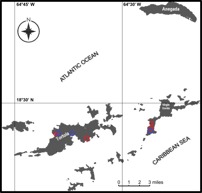
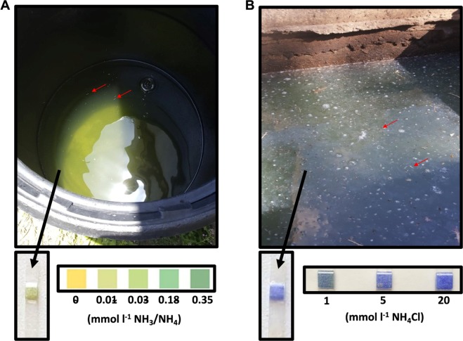
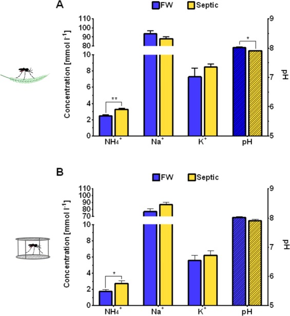
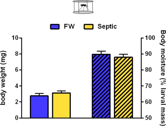
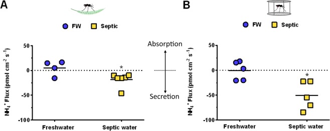
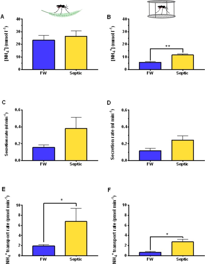
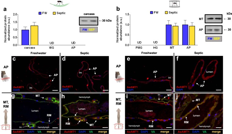
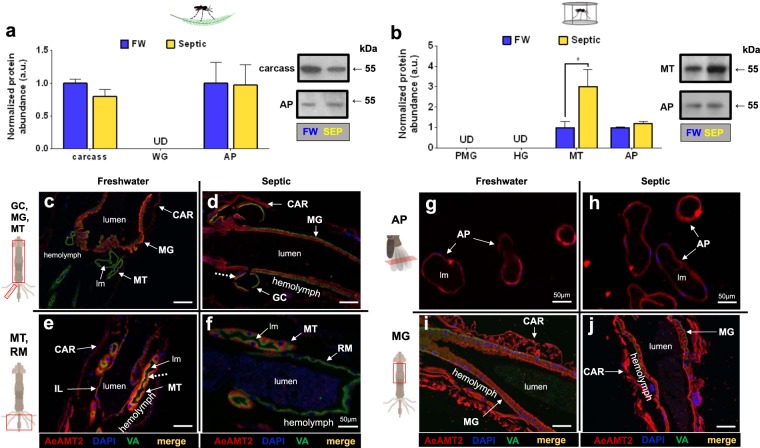
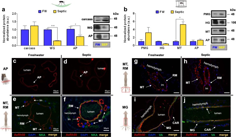
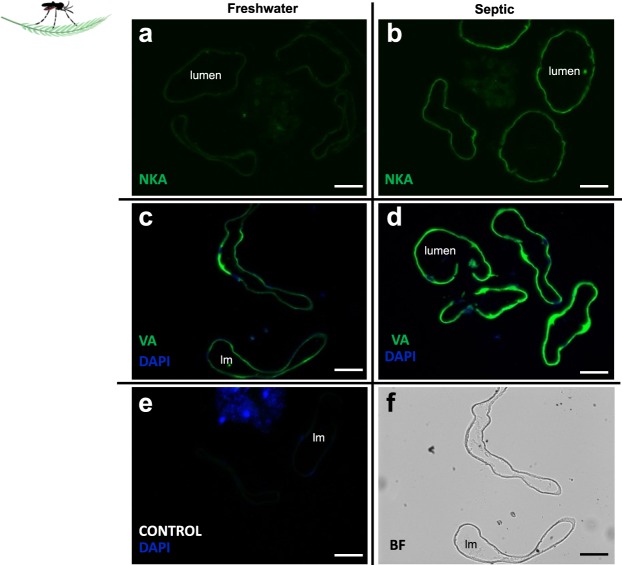
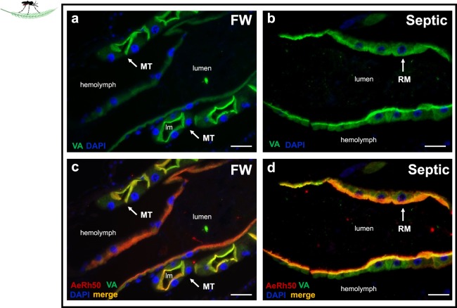
Larvae of the disease vector mosquito, Aedes aegypti (L.) readily develop in ammonia rich sewage in the British Virgin Islands. To understand how the larvae survive in ammonia levels that are lethal to most animals, an examination of ammonia excretory physiology in larvae collected from septic-water and freshwater was carried out. A. aegypti larvae were found to be remarkably plastic in dealing with high external ammonia through the modulation of NH4+ excretion at the anal papillae, measured using the scanning ion-selective electrode technique (SIET), and NH4+ secretion in the primary urine by the Malpighian tubules when developing in septicwater. Ammonia transporters, Amt and Rh proteins, are expressed in ionoregulatory and excretory organs, with increases in Rh protein, Na+-K+-ATPase, and V-type-H+-ATPase expression observed in the Malpighian tubules, hindgut, and anal papillae in septic-water larvae. A comparative approach using laboratory A. aegypti larvae reared in high ammonia septic-water revealed similar responses to collected A. aegypti with regard to altered ammonia secretion and hemolymph ion composition. Results suggest that the observed alterations in excretory physiology of larvae developing in septic-water is a consequence of the high ammonia levels and that A. aegypti larvae may rely on ammonia transporting proteins coupled to active transport to survive in septic-water.


Conflict of interest statement

The authors declare no competing interests.

Figures

Figure 1
Figure 1
Map of the study area and sites of A. aegypti larvae collection within the British Virgin Islands. The location of Aedes aegypti larvae collected from septic tanks (6 septic tanks within 4 different areas, red circles) and artificial containers containing freshwater (5 artificial containers within 3 different areas, blue circles) used in the present study are indicated. A. aegypti larvae were collected from urbanized sites on the islands of Tortola and Virgin Gorda.
Figure 2
Figure 2
Representative images of a freshwater container and a septic tank containing raw sewage water each with actively breeding A. aegypti used in the present study. (A) Barrel containing freshwater (FW) and (B) septic tank with raw sewage water each containing live A. aegypti larvae (indicated by red arrows). Representative ammonia (NH3/NH4+) test strips (Tetra EasyStrips) from one FW site and one septic water site used to estimate ammonia levels are shown below each image (A,B) along with a control concentration gradient.
Figure 3
Figure 3
Hemolymph ion and pH levels of wild-collected and laboratory A. Aegypti larvae reared in freshwater (FW) and septic water (Septic). Ammonium (NH4+), sodium (Na+), potassium (K+) and pH (dashed bars) levels in the hemolymph of (A) wild A. aegypti larvae (n = 6–7 per group) and (B) laboratory A. aegypti larvae (n = 15–17 per group) reared in FW or Septic. Data shown as mean ± S.E.M. Asterisks indicate statistical significance (*p < 0.05; **p < 0.005) compared to FW control (Unpaired, two-tailed t-test; Adjusted p values shown; Holm-Sidak correction for multiple comparisons).
Figure 4
Figure 4
Mean body weight and total body moisture (dashed bars) of laboratory A. aegypti larvae reared in freshwater (FW) or septic water (Septic). Data shown as mean ± S.E.M (n = 9 for FW, n = 9 for Septic). [Unpaired, two-tailed t-test; p = 0.35 for body weight, p = 0.51 for body moisture].
Figure 5
Figure 5
Scanning ion-selective micro-electrode technique (SIET) measurements of NH4+ flux at the anal papillae of wild-collected and laboratory A. aegypti larvae reared in freshwater (FW) and septic water (Septic). (A) NH4+ flux at the anal papillae of wild A. aegypti larvae reared in FW or Septic (n = 4 for FW, n = 6 for Septic), and (B) NH4+ flux at the anal papillae of laboratory A. aegypti larvae reared in FW of Septic (n = 5 for FW and Septic), measured each in their respective freshwater or septic water baths. NH4+ flux for each individual animal is shown as a single point, with the mean flux for each group illustrated by a horizontal solid black line. Negative values indicate efflux, or excretion, and positive values indicate influx, or absorption from the external. Data shown as mean ± S.E.M. Asterisks indicate statistical significance (*p < 0.05) compared to FW control (Unpaired, two-tailed t-test).
Figure 6
Figure 6
Transepithelial fluid secretion rates, ammonium (NH4+) concentrations in the secreted fluid, and NH4+ transport rates of Malpighian tubules from wild-collected and laboratory A. aegypti larvae reared in freshwater (FW) or septic water (Septic). NH4+ concentrations in Malpighian tubule secreted fluid from (A) wild A. aegypti larvae and (B) laboratory A. aegypti larvae reared in FW and septic water. Transepithelial fluid secretion rate of the Malpighian tubules from (C) wild A. aegypti larvae and (D) laboratory A. aegypti larvae reared in FW and septic water. NH4+ transport rate by the Malpighian tubules from (E) wild A. aegypti larvae and (F) laboratory A. aegypti larvae reared in FW and septic water. Data shown as mean ± S.E.M (n = 5–7 wild larvae, n = 5 laboratory larvae). Asterisks indicate statistical significance (*p < 0.05; **p < 0.005) compared to FW control (Unpaired, two-tailed t-test).
Figure 7
Figure 7
AeAmt1 abundance and immunolocalization in the alimentary canal, anal papillae, and carcass of wild-collected and laboratory A. aegypti larvae reared in freshwater (FW) and septic water (Septic). (a) AeAmt1 abundance and representative Western blot (right panel) in the carcass of wild A. aegypti larvae (n = 3). (b) AeAmt1 abundance and representative Western blot (right panel) in the Malpighian tubules (MT) and anal papillae (AP) of laboratory A. aegypti larvae (n = 3 FW, n = 4 Septic). The abundance of AeAmt1 protein was normalized to total protein (Coomassie protein stain, not shown), and Septic values are expressed relative to the control FW group (assigned a value of 1). Data shown as mean ± S.E.M. [Unpaired, two-tailed t-test; p < 0.05]. Representative transverse sections of anal papillae (AP) showing AeAmt1 (red) immunostaining from (c) wild FW-reared larvae, (d) wild Septic-reared larvae, (e) laboratory FW-reared larvae and (f) laboratory Septic-reared larvae. Nuclei are labelled by DAPI (blue) staining. Representative cross sections of the Malpighian tubules (MT) and rectum (RM) showing AeAmt1 (red) immunostaining from (g) wild FW-reared larvae, (h) wild Septic-reared larvae, (i) laboratory FW-reared larvae and (j) laboratory Septic-reared larvae. Nuclei are labelled by DAPI (blue) staining. Immunostaining of V1 subunit of V-type H+-ATPase (VA) is green (gj). Co-localization of AeAmt1 with apical V1 subunit of V-type H+-ATPase is indicated (dashed arrows) in the MT and RM (merge, yellow). Control sections (primary antibodies omitted, not shown) were devoid of red and green staining. Illustrations of the alimentary canal and anal papillae of A. aegypti larvae to the left of each immunofluorescence image indicates the region of the cross or transverse section (red rectangles). Lumen, (lm); anal papillae (AP); rectum (RM). Scale bars: 50 µm (cj).
Figure 8
Figure 8
AeAmt2 abundance and immunolocalization in the alimentary canal, anal papillae, and carcass of wild-collected and laboratory A. aegypti larvae reared in freshwater (FW) and septic water (Septic). (a) AeAmt2 abundance and representative Western blots (right panel) in the carcass and anal papillae (AP) of wild A. aegypti larvae (n = 3). (b) AeAmt2 abundance and representative Western blots (right panel) in the Malpighian tubules (MT) and anal papillae (AP) of laboratory A. aegypti larvae (n = 3 FW, n = 4 Septic). The abundance of AeAmt2 protein was normalized to total protein (Coomassie protein stain, not shown), and Septic values are expressed relative to the control FW group (assigned a value of 1). Data shown as mean ± S.E.M. Asterisks indicate statistical significance (*p < 0.05) compared to FW control (Unpaired, two-tailed t-test). Representative cross sections of the carcass (CAR), gastric caecae (GC), anterior and posterior midgut (MG) and Malpighian tubules (MT) and rectum (RM) showing AeAmt2 (red) immunostaining from (ce) wild FW-reared larvae, (df) wild Septic-reared larvae. Representative cross sections of the anterior midgut (MG) and transverse sections of anal papillae (AP) showing AeAmt2 (red) immunostaining from (gi) laboratory FW-reared larvae and (hj) laboratory Septic-reared larvae. Nuclei are labelled by DAPI (blue) staining. Immunostaining of V1 subunit of V-type H+-ATPase (VA) is green (ch). Co-localization of AeAmt2 with V1 subunit of V-type H+-ATPase is indicated (dashed arrows) (merge, yellow). Control sections (primary antibodies omitted, not shown) were devoid of red and green staining. Illustrations of the alimentary canal and anal papillae of A. aegypti larvae to the left of each immunofluorescence image indicates the region of the cross or transverse section (red rectangles). Lumen, (lm); carcass (CAR), gastric caecae (GC), midgut (MG), Malpighian tubule (MT), anal papillae (AP); rectum (RM). Scale bars: 100 µm, unless specified.
Figure 9
Figure 9
Rh protein (AeRh50) abundance and immunolocalization in the alimentary canal, anal papillae, and carcass of wild-collected and laboratory A. aegypti larvae reared in freshwater (FW) and septic water (Septic). (a) AeRh50 abundance and representative Western blots (right panel) in the epidermis, whole gut (WG), and anal papillae (AP) of wild A. aegypti larvae (n = 3). (b) AeRh50 abundance and representative Western blots (right panel) in the posterior midgut (PMG), hindgut (HG), Malpighian tubules (MT) and anal papillae (AP) of laboratory A. aegypti larvae (n = 3 FW, n = 4 Septic). The abundance of AeRh50 protein was normalized to total protein (Coomassie protein stain, not shown), and Septic values are expressed relative to the control FW group (assigned a value of 1). Data shown as mean ± S.E.M. Asterisks indicate statistical significance (*p < 0.05; ***p < 0.001) compared to FW control (Unpaired, two-tailed t-test). Representative transverse and cross sections of the anal papillae (AP), Malpighian tubules (MT) and rectum (RM) showing AeRh50 (red) immunostaining from (ce) wild FW-reared larvae, (df) wild Septic-reared larvae. Representative cross sections of the posterior midgut (MG), Malpighian tubules, rectum (RM), and carcass (CAR) showing AeRh50 (red) immunostaining from (gi) laboratory FW-reared larvae and (hj) laboratory Septic-reared larvae. Nuclei are labelled by DAPI (blue) staining. Immunostaining of Na+-K+-ATPase (NKA) (e,f) and the V1 subunit of V-type H+-ATPase (VA) (ij) are shown in green. Co-localization of AeRh50 with V1 subunit of V-type H+-ATPase is indicated (dashed arrows) (merge, yellow). Control sections (primary antibodies omitted, not shown) were devoid of red and green staining. Illustrations of the alimentary canal and anal papillae of A. aegypti larvae to the left of each immunofluorescence image indicates the region of the cross or transverse section (red rectangles). Lumen (lm), midgut (MG), Malpighian tubule (MT), anal papillae (AP); rectum (RM). Scale bars: 100 µm, unless specified.
Figure 10
Figure 10
Na+-K+-ATPase (NKA) and V-type H+-ATPase (VA) immunolocalization in the anal papillae (AP) of wild-collected A. aegypti larvae reared in freshwater (FW) and septic water (Septic). NKA immunostaining (green) of representative transverse sections of the AP from (a) FW-reared larvae and (b) Septic-reared larvae. VA immunostaining (green) of representative transverse sections of the AP from (c) FW-reared larvae and (d) Septic-reared larvae. (e) Control sections of AP (CONTROL, primary antibody omitted). DAPI staining of nuclei is in blue. (f) Representative bright field (BF) image of AP transverse sections in C. Scale bars: 50 µm. Lumen (lm), anal papillae (AP).
Figure 11
Figure 11
V-type H+-ATPase (VA) immunolocalization in the rectum (RM) of wild-collected A. aegypti larvae reared in freshwater (FW) and septic water (Septic). VA immunostaining (green) in representative paraffin-embedded cross sections of the (a) RM and Malpighian tubules (MT) from FW-reared wild A. aegypti larvae and (b) RM of septic-reared wild A. aegypti larvae. Nuclei are labelled with DAPI (blue). (c) cross section of MT and RM from (a) with apical AeRh50 staining (red) showing colocalization of with VA in MT (merge, yellow) in FW larvae and (d) cross section of RM corresponding to (b) showing co-localization (merge, yellow) of VA with apical AeRh50 staining (red) in septic larvae. Scale bars: 50 µm. Lumen (lm); rectum (RM), Malpighian tubule (MT).

Similar articles

Cited by

References

    1. Bhatt S, et al. The global distribution and burden of dengue. Nature. 2013;496:504–507. doi: 10.1038/nature12060. - DOI - PMC - PubMed
    1. Gubler DJ. Resurgent Vector-Borne Diseases as a Global Health Problem. Emerg. Infect. Dis. 1998;4:442–450. doi: 10.3201/eid0403.980326. - DOI - PMC - PubMed
    1. Burke RL, Barrera R, Lewis M, Kluchinsky T, Claborn D. Septic tanks as larval habitats for the mosquitoes Aedes aegypti and Culex quinquefasciatus in Playa-Playita, Puerto Rico. Med. Vet. Entomol. 2010;24:117–123. doi: 10.1111/j.1365-2915.2010.00864.x. - DOI - PubMed
    1. Irving‐Bell RJ, Okoli EI, Diyelong DY, Lyimo EO, Onyia OC. Septic tank mosquitoes: competition between species in central Nigeria. Med. Vet. Entomol. 1987;1:243–250. doi: 10.1111/j.1365-2915.1987.tb00350.x. - DOI - PubMed
    1. Lam WK, Dharmaraj D. A survey on mosquitoes breeding in septic tanks in several residential areas around Ipoh municipality. Med. J. Malaysia. 1982;37:114–123. - PubMed

Publication types

MeSH terms