. 2019 Dec 13;9(1):19116.
doi: 10.1038/s41598-019-55655-0.

New Class of Crosslinker-Free Nanofiber Biomaterials from Hydra Nematocyst Proteins

Affiliations

New Class of Crosslinker-Free Nanofiber Biomaterials from Hydra Nematocyst Proteins

Theresa Bentele et al. Sci Rep. .

Abstract

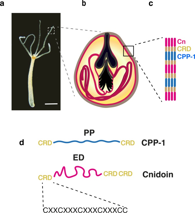
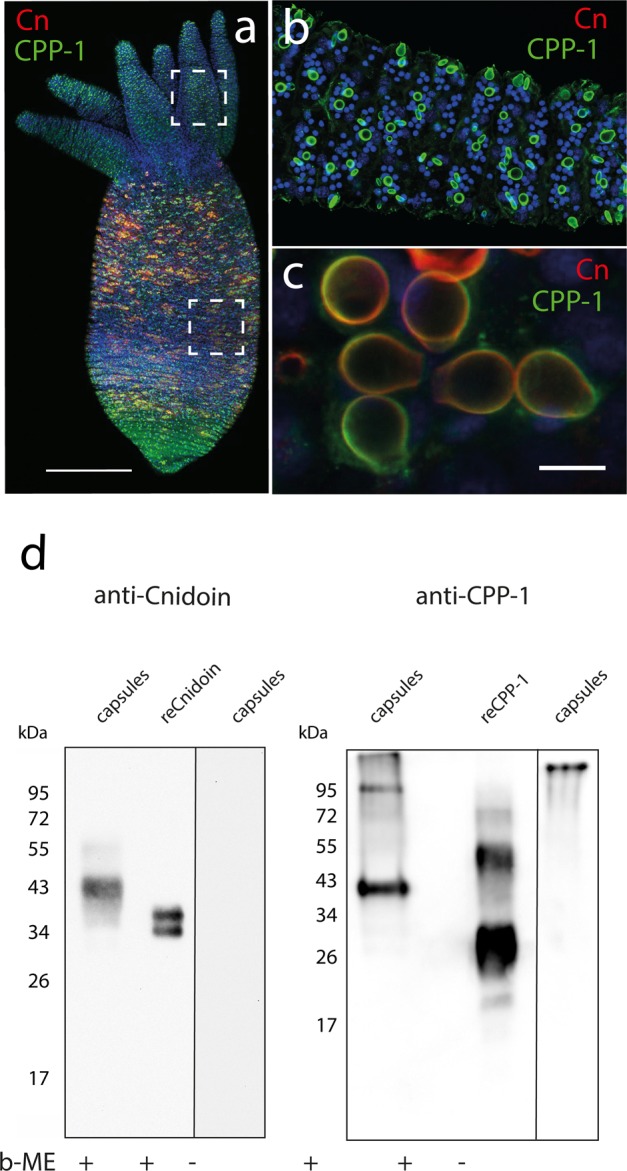
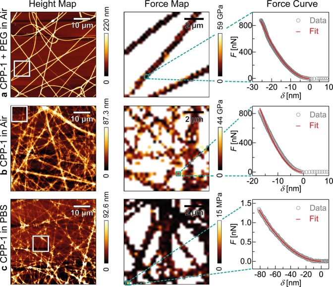
Nematocysts, the stinging organelles of cnidarians, have remarkable mechanical properties. Hydra nematocyst capsules undergo volume changes of 50% during their explosive exocytosis and withstand osmotic pressures of beyond 100 bar. Recently, two novel protein components building up the nematocyst capsule wall in Hydra were identified. The cnidarian proline-rich protein 1 (CPP-1) characterized by a "rigid" polyproline motif and the elastic Cnidoin possessing a silk-like domain were shown to be part of the capsule structure via short cysteine-rich domains that spontaneously crosslink the proteins via disulfide bonds. In this study, recombinant Cnidoin and CPP-1 are expressed in E. coli and the elastic modulus of spontaneously crosslinked bulk proteins is compared with that of isolated nematocysts. For the fabrication of uniform protein nanofibers by electrospinning, the preparative conditions are systematically optimized. Both fibers remain stable even after rigorous washing and immersion into bulk water owing to the simultaneous crosslinking of cysteine-rich domains. This makes our nanofibers clearly different from other protein nanofibers that are not stable without chemical crosslinkers. Following the quantitative assessment of mechanical properties, the potential of Cnidoin and CPP-1 nanofibers is examined towards the maintenance of human mesenchymal stem cells.


Conflict of interest statement

The authors declare no competing interests.

Figures

Figure 1
Figure 1
(a) Bright field image of a Hydra polyp (scale bar: 500 µm). (b) Schematic representation of a stenothele-type nematocyst with a large stylet apparatus and a coiled tubule inside of the hollow capsule body. (c) The nematocyst capsule wall consists of CPP-1 and Cnidoin (Cn), linked via cysteine-rich domains (CRDs). (d) CPP-1 has a “rigid” polyproline domain (PP) flanked by two CRD units, while Cnidoin consists of an “elastic”, silk-like domain (ED) flanked by CRD units. Each CRD unit has six cysteine residues in a conserved pattern (X denotes a non-cysteine residue).
Figure 2
Figure 2
(a) Immunofluorescence image of a Hydra polyp stained with CPP-1 and Cnidoin antibodies; cell nuclei (blue), CPP-1 (green), and Cnidoin (red). (b) Mature capsules in tentacles showed only CPP-1 signals. (c) Zoom-in images of capsules in the gastric region indicated co-localization of CPP-1 and Cnidoin in nematocyst walls. (d) Western blot analysis of CPP-1 and Cnidoin in isolated nematocysts and after recombinant expression in E. coli (reCPP-1, reCnidoin). (+) and (−) indicate the presence or absence of β−mercaptoethanol (β−ME) in the sample buffer. Uncropped images of gels are presented in Figure S4.
Figure 3
Figure 3
(a) Left: SEM image of isolated, partly discharged nematocysts. Right: Bright field microscopy image of an isolated discharged stenothele. The black triangle shadow corresponds to the AFM cantilever. (b) Height map of the discharged nematocyst collected from the red square in (a) (17 × 17 µm2). (c) A typical force-indentation curve measured on the nematocyst at the position indicated by the red square in (b) (1.1 × 1.1 µm2). The force-indentation data (gray circles) was fitted with the Bilodeau model for pyramidal tips (red curve).
Figure 4
Figure 4
AFM measurements of electrospun reCPP-1 fibers. First, a reCPP-1:PEG (1:1) mixture was electrospun and characterized in air (a). Second, the reCPP-1:PEG fibers were washed by water, and the remaining reCPP-1 fibers were characterized in air (b), as well as in PBS (c). Each dataset consists of height maps (left column), force maps (middle column), and characteristic force-indentation curves (right column) fitted with the Bilodeau model (red curve).

References

    1. Beckmann A, Özbek S. The nematocyst: a molecular map of the cnidarian stinging organelle. Int. J. Dev. Biol. 2012;56:577–582. doi: 10.1387/ijdb.113472ab. - DOI - PubMed
    1. Bode HR, Flick KM. Distribution and dynamics of nematocyte populations in Hydra attenuata. J. Cell Sci. 1976;21:15–34. - PubMed
    1. Chapman GB, Tilney LG. Cytological studies of the nematocysts of Hydra. I. Desmonemes, isorhizas, cnidocils, and supporting structures. J. Biophys. Biochem. Cytol. 1959;5:69–78. doi: 10.1083/jcb.5.1.69. - DOI - PMC - PubMed
    1. Chapman GB, Tilney LG. Cytological studies of the nematocysts of Hydra. II. The stenoteles. J. Biophys. Biochem. Cytol. 1959;5:79–84. doi: 10.1083/jcb.5.1.79. - DOI - PMC - PubMed
    1. Holstein TW. A view to kill. BMC Biol. 2012;10:18. doi: 10.1186/1741-7007-10-18. - DOI - PMC - PubMed

Publication types

MeSH terms