Review
. 2020 Feb;16(2):81-90.
doi: 10.1038/s41574-019-0286-3. Epub 2019 Dec 13.

The role of macrophages in obesity-associated islet inflammation and β-cell abnormalities

Affiliations
Review

The role of macrophages in obesity-associated islet inflammation and β-cell abnormalities

Wei Ying et al. Nat Rev Endocrinol. 2020 Feb.

Abstract

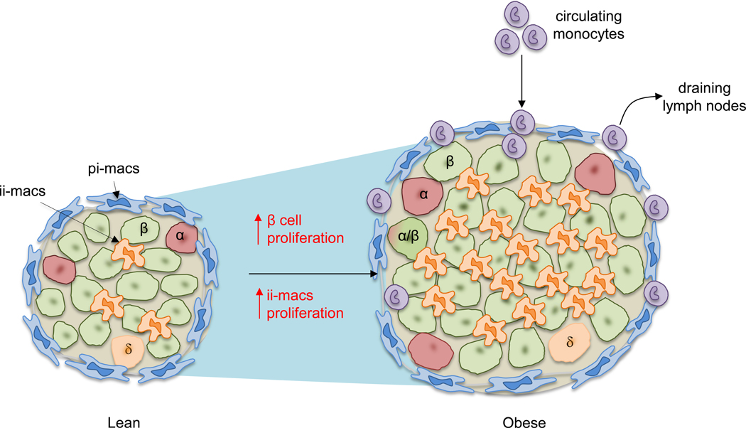
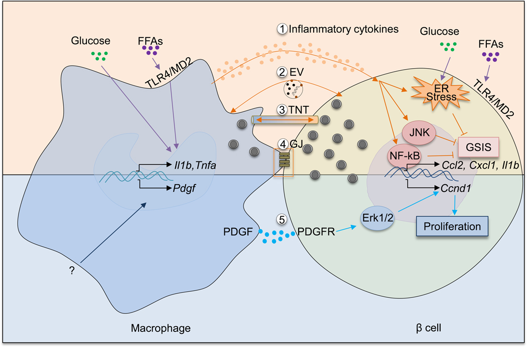
Chronic, unresolved tissue inflammation is a well-described feature of obesity, type 2 diabetes mellitus (T2DM) and other insulin-resistant states. In this context, adipose tissue and liver inflammation have been particularly well studied; however, abundant evidence demonstrates that inflammatory processes are also activated in pancreatic islets from obese animals and humans with obesity and/or T2DM. In this Review, we focus on the characteristics of immune cell-mediated inflammation in islets and the consequences of this with respect to β-cell function. In contrast to type 1 diabetes mellitus, the dominant immune cell type causing inflammation in obese and T2DM islets is the macrophage. The increased macrophage accumulation in T2DM islets primarily arises through local proliferation of resident macrophages, which then provide signals (such as platelet-derived growth factor) that drive β-cell hyperplasia (a classic feature of obesity). In addition, islet macrophages also impair the insulin secretory capacity of β-cells. Through these mechanisms, islet-resident macrophages underlie the inflammatory response in obesity and mechanistically participate in the β-cell hyperplasia and dysfunction that characterizes this insulin-resistant state. These findings point to the possibility of therapeutics that target islet inflammation to elicit beneficial effects on β-cell function and glycaemia.


Conflict of interest statement

DECLARATION OF INTERESTS

The authors have no conflicts of interests to declare.

Figures

Figure 1.
Figure 1.. Macrophages dominate obesity-associated islet inflammation.
Illustrated here is a comparison of mouse islets between lean and obese conditions. In lean mice, two major populations of macrophages can be detected based on their anatomical distributions: peri-islet macrophages (pi-macs) (F4/80hi CD11c-) and intra-islet macrophages (ii-macs) (F4/80lo CD11chi). Both are islet-resident cells. In contrast, in obesity, the size of islet is increased due to increased beta cell replication and cell size. Multiple studies have demonstrated the increase of islet macrophages. However, different mechanisms have been proposed to explain obesity-associated macrophage accumulation in the islet. In one model, stressed beta cells recruit circulating monocytes which differentiate into pro-inflammatory macrophages after infiltrating into the islets. This model has been challenged by other studies showing that even though monocytes can be detected in the pancreas, they do not infiltrate into the islets. Instead, the accumulation of intra-islet macrophages is caused by local proliferation of resident macrophages.
Figure 2.
Figure 2.. Interactions of islet macrophages and beta cells in obesity.
An increasing body of evidence supports the idea that islet macrophages influence beta cells in multiple ways. In obesity, elevated levels of glucose and free fatty acids can induce a pro-inflammatory phenotype of islet macrophages. As a result, macrophages produce increased amounts of proinflammatory cytokines such as IL-1β and TNF-α. These cytokines activate NF-κB and JNK pathways in beta cells and also exacerbate ER stress. Synergistically, these responses dampen beta cell GSIS. In addition to inflammatory cytokines, other mechanisms involving macrophage-mediated beta cell dysfunction exist. There mechanisms include: extracellular vesicles (EV) containing insulin released by beta cells and phagocytosed by islet macrophages; the formation of tunneling nanotubes (TNT) or gap junctions (GJ) between macrophages and beta cells allowing for bidirectional exchange of cellular contents. Obesity increases PDGF expression in islet macrophages via unclear mechanisms. Through PDGFR expressed in beta cells, PDGF promotes beta cell proliferation by activating downstream Erk signaling and inducing cell cycle gene (e.g., Ccnd1) expression.

References

    1. da Rocha Fernandes J et al. IDF Diabetes Atlas estimates of 2014 global health expenditures on diabetes. Diabetes Res Clin Pract 117, 48–54, doi:10.1016/j.diabres.2016.04.016 (2016). - DOI - PubMed
    1. Saisho Y Importance of Beta Cell Function for the Treatment of Type 2 Diabetes. J Clin Med 3, 923–943, doi:10.3390/jcm3030923 (2014). - DOI - PMC - PubMed
    1. Kitamura T The role of FOXO1 in beta-cell failure and type 2 diabetes mellitus. Nat Rev Endocrinol 9, 615–623, doi:10.1038/nrendo.2013.157 (2013). - DOI - PubMed
    1. Donath MY & Shoelson SE Type 2 diabetes as an inflammatory disease. Nat Rev. Immunol 11, 98–107, doi:nri2925 [pii];10.1038/nri2925 [doi] (2011). - DOI - PubMed
    1. Lackey DE & Olefsky JM Regulation of metabolism by the innate immune system. Nat Rev Endocrinol 12, 15–28, doi:10.1038/nrendo.2015.189 (2016). - DOI - PubMed

Publication types