

Cardiac T2 * mapping: Techniques and clinical applications
- PMID: 31837078
- PMCID: PMC7687175
- DOI: 10.1002/jmri.27023
Cardiac T2 * mapping: Techniques and clinical applications
Abstract
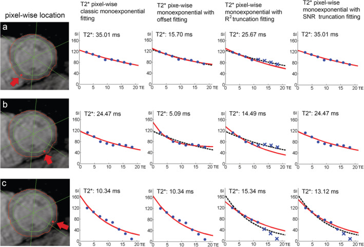
Cardiac T2 * mapping is a noninvasive MRI method that is used to identify myocardial iron accumulation in several iron storage diseases such as hereditary hemochromatosis, sickle cell disease, and β-thalassemia major. The method has improved over the years in terms of MR acquisition, focus on relative artifact-free myocardium regions, and T2 * quantification. Several improvement factors involved include blood pool signal suppression, the reproducibility of T2 * measurement as affected by scanner hardware, and acquisition software. Regarding the T2 * quantification, improvement factors include the applied curve-fitting method with or without truncation of the signals acquired at longer echo times and whether or not T2 * measurement focuses on multiple segmental regions or the midventricular septum only. Although already widely applied in clinical practice, data processing still differs between centers, contributing to measurement outcome variations. State of the art T2 * measurement involves pixelwise quantification providing better spatial iron loading information than region of interest-based quantification. Improvements have been proposed, such as on MR acquisition for free-breathing mapping, the generation of fast mapping, noise reduction, automatic myocardial contour delineation, and different T2 * quantification methods. This review deals with the pro and cons of different methods used to quantify T2 * and generate T2 * maps. The purpose is to recommend a combination of MR acquisition and T2 * mapping quantification techniques for reliable outcomes in measuring and follow-up of myocardial iron overload. The clinical application of cardiac T2 * mapping for iron overload's early detection, monitoring, and treatment is addressed. The prospects of T2 * mapping combined with different MR acquisition methods, such as cardiac T1 mapping, are also described. Level of Evidence: 4 Technical Efficacy Stage: 5 J. Magn. Reson. Imaging 2019.
Keywords: T2* techniques; cardiac T2* mapping; cardiac iron overload; magnetic resonance imaging.
© 2019 The Authors. Journal of Magnetic Resonance Imaging published by Wiley Periodicals, Inc. on behalf of International Society for Magnetic Resonance in Medicine.
Figures


References
-
- Aydinok Y, Porter JB, Piga A, et al. Prevalence and distribution of iron overload in patients with transfusion‐dependent anemias differs across geographic regions: Results from the CORDELIA study. Eur J Haematol 2015;95:244–253. - PubMed
-
- Chen X, Zhang Z, Zhong J, et al. MRI assessment of excess cardiac iron in thalassemia major: When to initiate? J Magn Reson Imaging 2015;42:737–745. - PubMed
-
- Eghbali A, Shokri P, Afzal RR, Bagheri B. A 1‐year randomized trial of deferasirox alone versus deferasirox and deferoxamine combination for the treatment of iron overload in thalassemia major. Transfus Apher Sci 2019. [Epub ahead of print]. - PubMed
Publication types
MeSH terms
LinkOut - more resources
Full Text Sources
