. 2020 Apr:212:7-16.
doi: 10.1016/j.ajo.2019.11.031. Epub 2019 Dec 16.

Retinal Vessel Density in Exudative and Nonexudative Age-Related Macular Degeneration on Optical Coherence Tomography Angiography

Affiliations

Retinal Vessel Density in Exudative and Nonexudative Age-Related Macular Degeneration on Optical Coherence Tomography Angiography

Sophie C Lee et al. Am J Ophthalmol. 2020 Apr.

Abstract

Purpose: Although the choroid contributes to the pathogenesis of age-related macular degeneration (AMD), the role of retinal perfusion is unclear. We sought to compare retinal vascular measurements between eyes with nonexudative and exudative AMD using optical coherence tomography angiography (OCT-A).

Design: Retrospective, cross-sectional study.

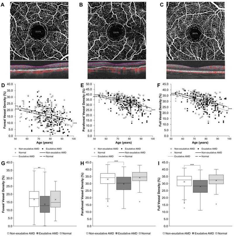
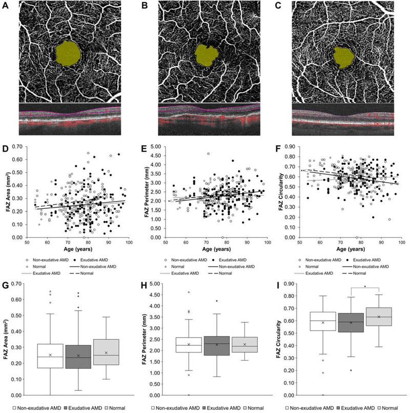
Methods: OCT-A images were analyzed from 310 eyes of 182 patients (mean age ± standard deviation [SD], 78.8 ± 8.8 years) with nonexudative (54.2%) and exudative (45.8%) AMD to measure retinal vessel density (VD) from the superficial capillary plexus in the foveal, parafoveal, and full macular regions and foveal avascular zone (FAZ) area, perimeter, and circularity. Multivariate regressions were used to compare nonexudative and exudative AMD eyes and the impact of anti-vascular endothelial growth factor (anti-VEGF) treatments or geographic atrophy (GA).

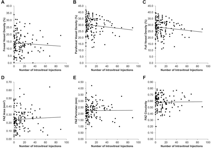
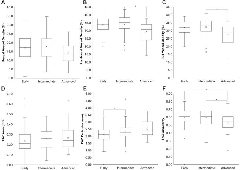
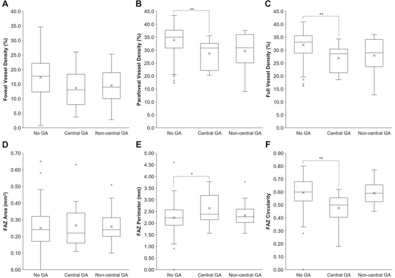
Results: In eyes with AMD, VD decreases with age in the foveal (β = -0.211, P < .001), parafoveal (β = -0.305, P < .001), and full macular regions (β = -0.295, P < .001). Eyes with exudative AMD demonstrated lower VD, especially in the parafoveal (29.8% ± 6.3% vs 33.0% ± 5.7%, P < .001) and full regions (27.9% ± 6.2% vs 31.2% ± 5.5%, P < .001) compared with nonexudative AMD. There were no differences in FAZ area, perimeter, or circularity between the 2 groups (P = .503-.907). In eyes with exudative AMD, previous anti-VEGF treatments did not impact retinal vascular measurements (P = .324-.986). Nonexudative AMD severity and presence of central GA also impacted retinal VD and FAZ morphology.

Conclusions: Retinal VD is decreased in eyes with exudative AMD compared with nonexudative AMD but is unaffected by anti-VEGF treatments, suggesting a retinal vascular contribution to the pathogenesis of AMD.


Figures

Figure 1.
Figure 1.. Retinal Vessel Density in Non-exudative & Exudative Age-Related Macular Degeneration.
Representative optical coherence tomography-angiography (OCT-A) and spectral-domain OCT scans of eyes with non-exudative (A) and exudative (B) age-related macular degeneration (AMD) and a normal control (C) with grid overlay demonstrating the central fovea, parafoveal inner ring, and full 3 mm macular region for vessel density (VD) measurements. Scatterplots with linear regression trendlines showing foveal (D), parafoveal (E), and full (F) VD in eyes with non-exudative and exudative AMD and normal eyes across different patient ages. Box-and- whisker plots comparing foveal (G), parafoveal (H), and full (I) VD in eyes with non-exudative and exudative AMD and normal eyes. **, P<0.01; ***, P<0.001
Figure 2.
Figure 2.. FAZ Measurements in Non-exudative and Exudative Age-Related Macular Degeneration.
Representative OCT-A and spectral-domain OCT scans of eyes with non-exudative (A) and exudative (B) AMD and a normal control (C) with foveal avascular zone (FAZ) area highlighted. Scatterplots with linear regression trendlines showing FAZ area (D), perimeter (E), and circularity (F) in eyes with non-exudative and exudative AMD and normal eyes across different patient ages. Box-and-whisker plots comparing FAZ area (G), perimeter (H), and circularity (I) in eyes with non-exudative and exudative AMD and normal eyes. *, P<0.05; **, P<0.01
Figure 3.
Figure 3.. Retinal Vascular Measurements after Anti-Vascular Endothelial Growth Factor Treatments.
Scatterplots with trendlines showing the relationship between the number of intravitreal anti-vascular endothelial growth factor (anti-VEGF) injections and foveal (A), parafoveal (B), and full (C) VD, as well as FAZ area (D), perimeter (E), and circularity (F) in eyes with exudative AMD.
Figure 4.
Figure 4.. Retinal Vascular Measurements in Non-exudative Age-Related Macular Degeneration.
Box-and-whisker plots comparing foveal (A), parafoveal (B), and full (C) VD, as well as FAZ area (D), perimeter (E), and circularity (F) in eyes with early, intermediate, or advanced non-exudative AMD. *, P<0.05
Figure 5.
Figure 5.. Retinal Vascular Measurements in Geographic Atrophy.
Box-and-whisker plots comparing foveal (A), parafoveal (B), and full (C) VD, as well as FAZ area (D), perimeter (E), and circularity (F) in eyes with non-exudative AMD and no geographic atrophy (GA), central GA, or non-central GA. *, P<0.05; **, P<0.01

References

    1. Al-Zamil WM, Yassin SA. Recent developments in age-related macular degeneration: a review. Clin Interv Aging 2017;12:1313–1330. - PMC - PubMed
    1. Shaw PX, Stiles T, Douglas C, et al. Oxidative stress, innate immunity, and age-related macular degeneration. AIMS Mol Sci 2016;3(2):196–221. - PMC - PubMed
    1. Jarrett SG, Boulton ME. Consequences of oxidative stress in age-related macular degeneration. Mol Aspects Med 2012;33(4):399–417. - PMC - PubMed
    1. Ferrington DA, Sinha D, Kaarniranta K. Defects in retinal pigment epithelial cell proteolysis and the pathology associated with age-related macular degeneration. Prog Retin Eye Res 2016;51:69–89. - PMC - PubMed
    1. Handa JT, Cano M, Wang L, Datta S, Liu T. Lipids, oxidized lipids, oxidation-specific epitopes, and Age-related Macular Degeneration. Biochim Biophys Acta Mol Cell Biol Lipids 2017;1862(4):430–440. - PMC - PubMed

Publication types