. 2020 Jan:310:106664.
doi: 10.1016/j.jmr.2019.106664. Epub 2019 Nov 28.

Proton-detected polarization optimized experiments (POE) using ultrafast magic angle spinning solid-state NMR: Multi-acquisition of membrane protein spectra

Affiliations

Proton-detected polarization optimized experiments (POE) using ultrafast magic angle spinning solid-state NMR: Multi-acquisition of membrane protein spectra

T Gopinath et al. J Magn Reson. 2020 Jan.

Abstract

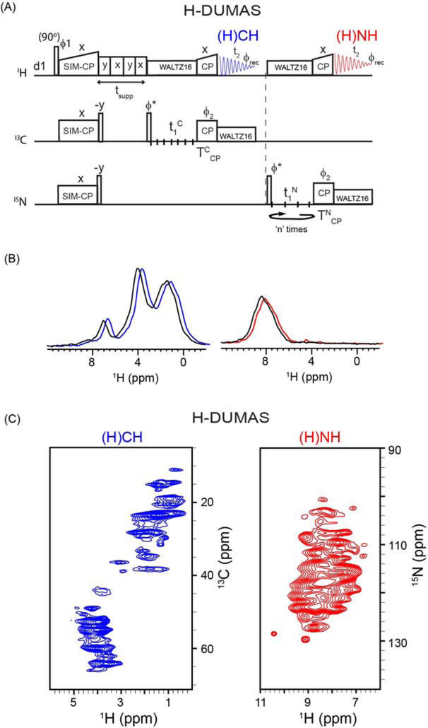
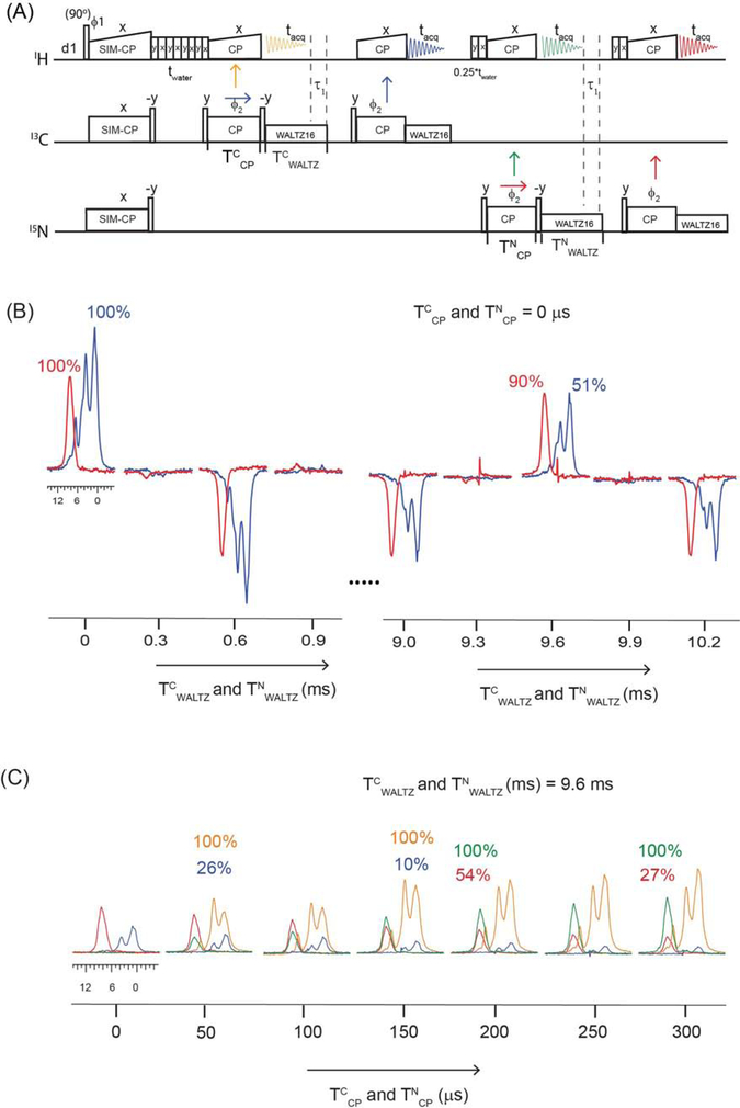
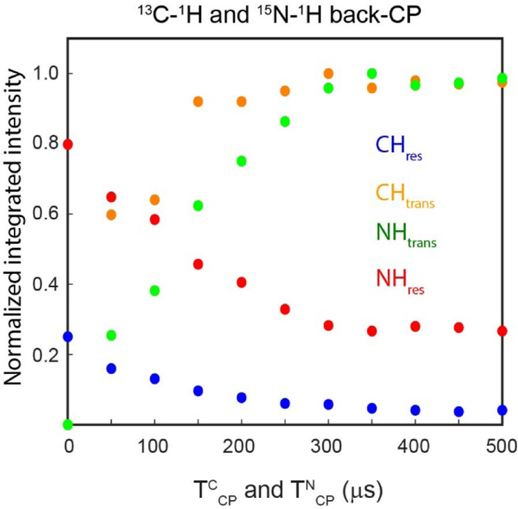
Proton-detected solid-state NMR (ssNMR) spectroscopy has dramatically improved the sensitivity and resolution of fast magic angle spinning (MAS) methods. While relatively straightforward for fibers and crystalline samples, the routine application of these techniques to membrane protein samples is still challenging. This is due to the low sensitivity of these samples, which require high lipid:protein ratios to maintain the structural and functional integrity of membrane proteins. We previously introduced a family of novel polarization optimized experiments (POE) that enable to make the best of nuclear polarization and obtain multiple-acquisitions from a single pulse sequence and one receiver. Here, we present the 1H-detected versions of POE using ultrafast MAS ssNMR. Specifically, we implemented proton detection into our three main POE strategies, H-DUMAS, H-MEIOSIS, and H-MAeSTOSO, achieving the acquisition of up to ten different experiments using a single pulse sequence. We tested these experiments on a model compound N-Acetyl-Val-Leu dipeptide and applied to a six transmembrane acetate transporter, SatP, reconstituted in lipid membranes. These new methods will speed up the spectroscopy of challenging biomacromolecules such as membrane proteins.

Keywords: H-DUMAS; H-MAeSTOSO; H-MEIOSIS; Membrane proteins; Multi-acquisition; Polarization optimized experiments (POE); SIM-CP; Solid-state NMR; Ultra-fast magic angle spinning.


Conflict of interest statement

The authors declare that they have no known competing financial interests or personal relationships that could have appeared to influence the work reported in this paper.

Figures

Figure 1:
Figure 1:
(A) CP and SIM-CP pulse sequences used for quantifying relative signal intensities. For SIM-CP, 13C and 15N spectra were acquired in separate experiments with a single receiver. (B) and (C) Comparison of CP and SIM-CP on SatP membrane protein obtained from integrated intensities of 13C and 15N spectra acquired at 65 kHz and 12 kHz MAS rates, respectively.
Figure 2:
Figure 2:
(A) Pulse sequence for two-dimensional H-DUMAS (proton detected DUMAS) experiment for simultaneous acquisition of 13C- and 15N-edited experiments, (H)CH and (H)NH. (B) 1D (H)CH and (H)NH spectra of SatP membrane protein obtained from first increment (t1=0) of H-DUMAS (shown in blue and red), and the corresponding spectra (shown in black) obtained from single acquisition methods using 65 kHz MAS rate. For comparison the 1D spectra shown in blue and red are slightly shifted to the right. (C) Two-dimensional (H)CH and (H)NH spectra of SatP protein acquired using H-DUMAS pulse sequence with n=2 loops for 15N t1 evolution.
Figure 3:
Figure 3:
MEIOSIS experiment at fast MAS rates. (A) Pulse sequence for 1D MEIOSIS that records four polarization pathways (CCres, NCAtrans, NNres, and CANtrans) resulting from the first SPECIFIC-CP period τ. (B) Normalized integrated intensities of all four pathways measured for SatP protein by varying the τ mixing times. The polarization in each pathway is normalized with respect to initial value at τ=0. (C) 1D spectra of CCres, NCAtrans, NNres, and CANtrans (color coded accordingly) polarization pathways obtained at τ= 0 and 6.5 ms. (D) 1D spectra at τ=6.5 ms with corresponding intensities normalized with respect to highest intensity spectrum of CC polarization pathway.
Figure 4:
Figure 4:
(A) Two-dimensional H-MEIOSIS (proton detected MEIOSIS) pulse sequence for simultaneous acquisition of two pairs of 13C- and 15N-edited experiments, (H)CH, (H)N(CA)H, (H)NH and (H)CA(N)H. (B) 2D H-MEIOSIS spectra of SatP protein acquired at 65 kHz MAS rate using two loops (n=2) for 15N t1 evolution. A pair of 2D spectra in the 1st and 2nd acquisitions are obtained by adding and subtracting the two data sets recorded with ϕ=x and −x.
Figure 5:
Figure 5:
(A) Pulse sequence for evaluating the residual 13C and 15N polarization that remains on 13C and 15N at the end of back CP periods (TCCP and TNCP). (B) Membrane protein SatP spectra acquired in 2nd and 4th acquisition periods by varying TCWALTZ and TNWALTZ decoupling periods. (C) Transferred and residual polarization of 13C–1H and 15N–1H back CP periods measured by varying TCCP and TNCP back CP periods.
Figure 6:
Figure 6:
Normalized integrated intensities of transferred and residual polarization pathways of 13C–1H and 15N–1H back CP obtained from the spectra reported in Figure 5C using the pulse sequences of Figure 5A.
Figure 7:
Figure 7:
(A) Pulse sequence for the H-MAeSTOSO-4 experiment for simultaneous acquisition of (H)CHH, (H)NHH (RFDR=1.47 ms), H(N)HH (RFDR=0.74ms), and (H)NH experiments. Corresponding 2D spectra on SatP membrane protein are shown in (B). Suppression of 1H signals from water and lipids is obtained by using tsupp=200ms. Prior to 3rd and 4th acquisitions 0.25*tsupp (=50 ms) period was used for eliminating waters signals. (C) 2D spectra (H)NHH and (H)NH obtained from H-MAeSTOSO-4 pulse sequence in the absence of 0.25*tsupp period, showing the residual water signals.
Figure8:
Figure8:
(A) Pulse sequence for the H-MAeSTOSO-10 experiment for simultaneous acquisition of ten 2D spectra. (B) Application of H-MAeSTOSO-10 on the NAVL (N-acetyl-Val-Leu) dipeptide.

References

    1. Hu F; Luo W; Hong M, Mechanisms of proton conduction and gating in influenza M2 proton channels from solid-state NMR. Science 2010, 330 (6003), 505–8. - PMC - PubMed
    1. Quinn CM; Wang M; Fritz MP; Runge B; Ahn J; Xu C; Perilla JR; Gronenborn AM; Polenova T, Dynamic regulation of HIV-1 capsid interaction with the restriction factor TRIM5alpha identified by magic-angle spinning NMR and molecular dynamics simulations. Proc Natl Acad Sci U S A 2018, 115 (45), 11519–11524. - PMC - PubMed
    1. Zhou HX; Cross TA, Influences of membrane mimetic environments on membrane protein structures. Annu Rev Biophys 2013, 42, 361–92. - PMC - PubMed
    1. Baldus M, GPCR: Lock and key become flexible. Nat Chem Biol 2018, 14 (3), 201–202. - PubMed
    1. Fitzpatrick AW; Debelouchina GT; Bayro MJ; Clare DK; Caporini MA; Bajaj VS; Jaroniec CP; Wang L; Ladizhansky V; Muller SA; MacPhee CE; Waudby CA; Mott HR; De Simone A; Knowles TP; Saibil HR; Vendruscolo M; Orlova EV; Griffin RG; Dobson CM, Atomic structure and hierarchical assembly of a cross-beta amyloid fibril. Proc Natl Acad Sci U S A 2013, 110 (14), 5468–73. - PMC - PubMed

Publication types

LinkOut - more resources