Review
. 2020 Nov:66:116-128.
doi: 10.1016/j.semcancer.2019.11.007. Epub 2019 Dec 12.

Regulation of autophagy by canonical and non-canonical ER stress responses

Affiliations
Review

Regulation of autophagy by canonical and non-canonical ER stress responses

Monika Bhardwaj et al. Semin Cancer Biol. 2020 Nov.

Abstract

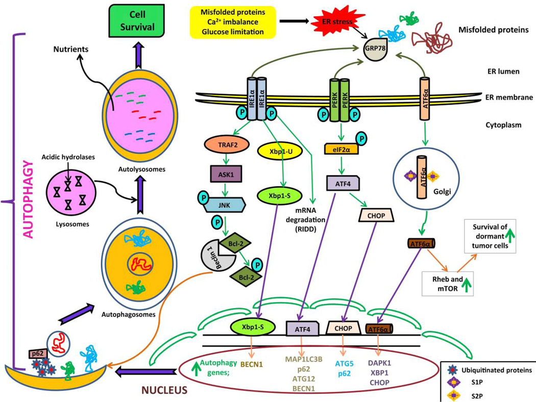
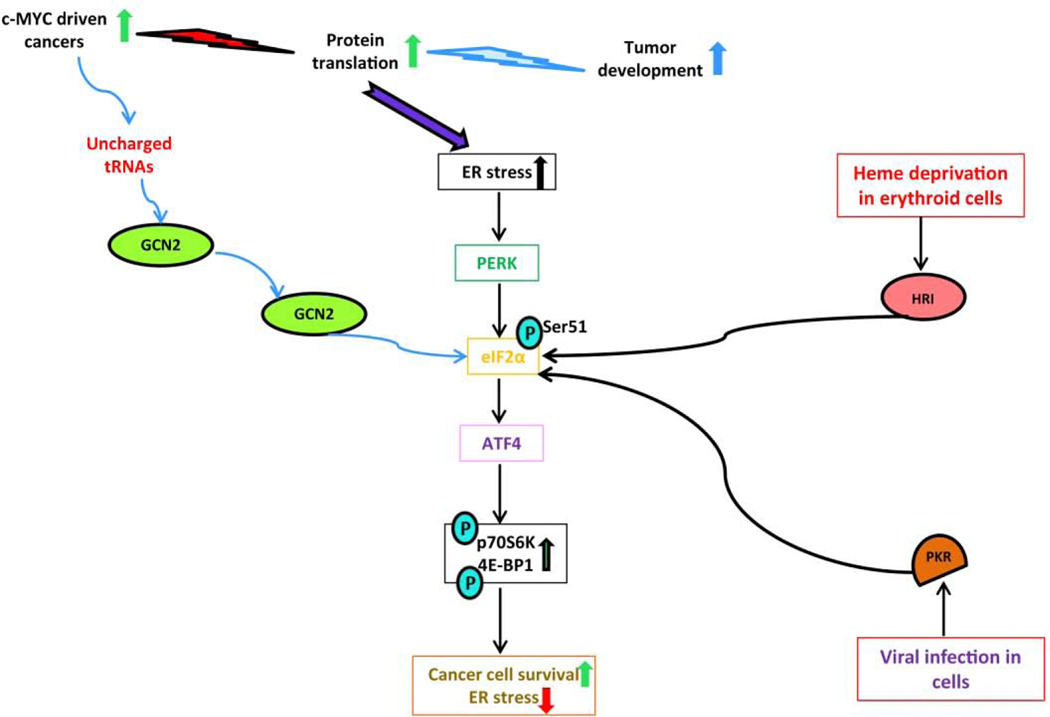
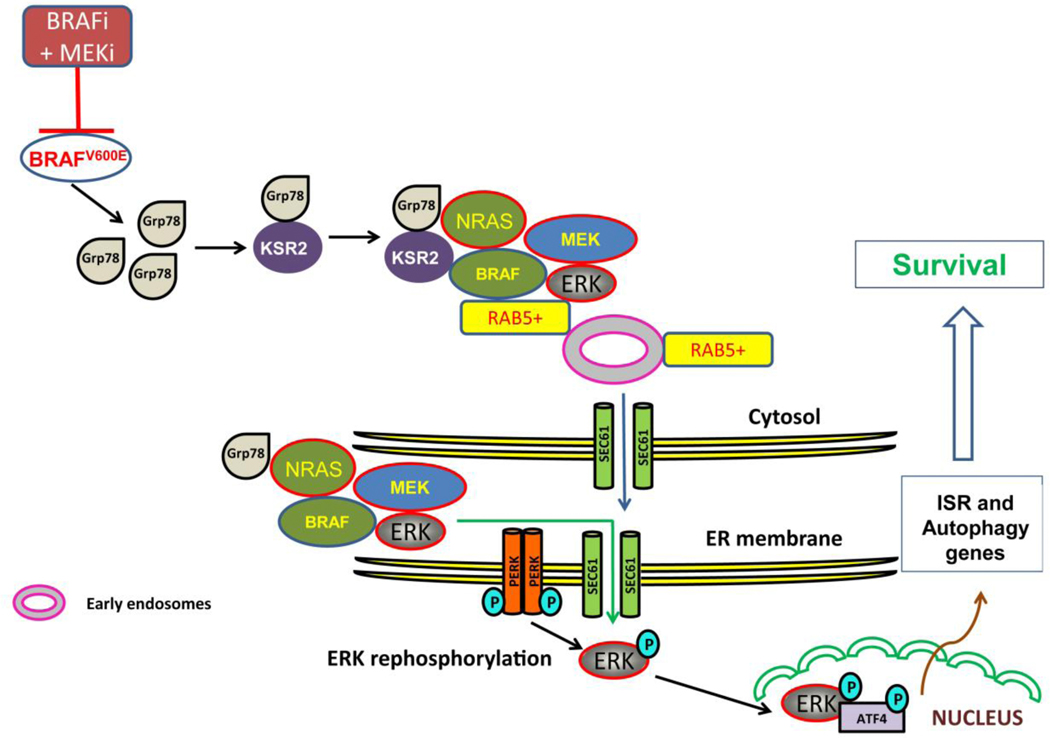
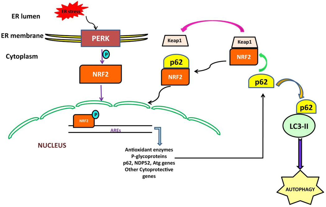
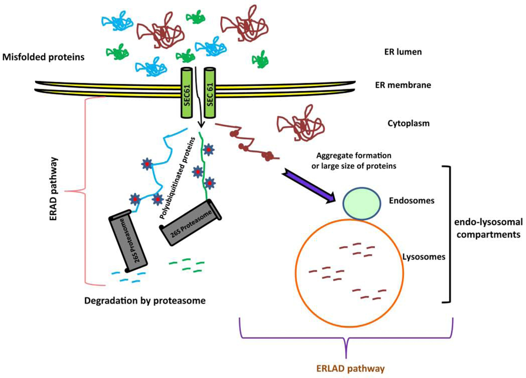
Cancer cells encounter numerous stresses that pose a threat to their survival. Tumor microenviroment stresses that perturb protein homeostasis can produce endoplasmic reticulum (ER) stress, which can be counterbalanced by triggering the unfolded protein response (UPR) which is considered the canonical ER stress response. The UPR is characterized by three major proteins that lead to specific changes in transcriptional and translational programs in stressed cells. Activation of the UPR can induce apoptosis, but also can induce cytoprotective programs such as autophagy. There is increasing appreciation for the role that UPR-induced autophagy plays in supporting tumorigenesis and cancer therapy resistance. More recently several new pathways that connect cell stresses, components of the UPR and autophagy have been reported, which together can be viewed as non-canonical ER stress responses. Here we review recent findings on the molecular mechanisms by which canonical and non-canonical ER stress responses can activate cytoprotective autophagy and contribute to tumor growth and therapy resistance. Autophagy has been identified as a druggable pathway, however the components of autophagy (ATG genes) have proven difficult to drug. It may be the case that targeting the UPR or non-canonical ER stress programs can more effectively block cytoprotective autophagy to enhance cancer therapy. A deeper understanding of these pathways could provide new therapeutic targets in cancer.

Keywords: Autophagy; Cancer; Canonical endoplasmic reticulum stress; Non-canonical endoplasmic reticulum stress; Unfolded protein response.


Conflict of interest statement

Conflict of interest statement: RKA is inventor on patents related to novel autophagy inhibitors and is founder of Pinpoint Therapeutics. He is on the scientific advisory board for Sprint Biosciences and Immunaccel, Inc.

Figures

Figure 1:
Figure 1:. The unfolded protein response (UPR) signaling mechanisms and autophagy.
Perturbances in endoplasmic reticulum (ER) environment due to misfolded proteins, Ca2+ imbalance, or glucose limitation causes ER stress. Following ER stress, the UPR is executed by three major proteins and their downstream effectors: inositol-requiring enzyme 1α (IRE1α), protein kinase R (PKR)-like endoplasmic reticulum kinase (PERK), and the activating transcription factor 6 α (ATF6α). The IRE1α arm of UPR produces spliced X box-binding protein 1 (XBP1-S) enhances the expression of Beclin-1 by binding with BECN1 gene. IRE1-dependent decay (RIDD) activation degrades UPR target mRNAs. Activated IRE1α forms a complex with tumor necrosis factorα (TNFα) receptor-associated factor 2 (TRAF2) and apoptosis signaling-regulating kinase (ASK1), which in turn activates c-Jun N-terminal kinase (JNK). The IRE1α-JNK arm mediates phosphorylation of B-cell lymphoma 2 (Bcl-2), which causes Beclin-1 dissociation and induction of autophagy. Autophagy is induced via expression of p62, autophagy regulated (ATG) 5, ATG12, and microtubule associated protein 1 light chain 3 beta (MAP1LC3B) genes by the activating transcription factor 4 (ATF4) transcription factor and CCAAT/enhancer binding protein homologous protein (CHOP) which is mediated by PERK arm of the UPR. The ATF6α arm of UPR is believed to induce autophagy by regulating the transcriptional levels of death-associated kinase 1 (DAPK1), CHOP and XBP1 genes. In addition, ATF6α upregulates Ras homology enriched in brain (Rheb) and mammalian target of rapamycin (mTOR) to promote survival of dormant tumor cells.
Figure 2:
Figure 2:. The Integrated stress response (ISR) in MYC-driven malignancies.
The ISR is activated by various intrinsic and extrinsic factors. Phosphorylation of eIF2α at Ser51 by double-stranded RNA-activated protein kinase (PKR), general control non-derepressible-2 (GCN2) and the heme-regulated inhibitor kinase (HRI) occurs during viral infection, amino-acid starvation and the heme-deprivation in erythroid cells respectively. During amino-acid starvation GCN2 is activated by uncharged tRNAs and phosphorylates eIF2α and triggers ATF4 induction in MYC driven cancers. Enhanced phosphorylation of p70S6K1 and 4E-BP1 abrogates ER stress and supports c-MYC driven cancer survival.
Figure 3:
Figure 3:. ER translocation of MAPK pathways and autophagy.
During targeted therapy with BRAF and MEK inhibition in BRAF mutant melanoma, a cytoplasmic pool of Grp78 binds to the scaffold protein KSR2 which recruits NRAS, BRAF, MEK and ERK. This multiprotein complex is shuttled to Rab5+ on early endosomes on ER membrane. MAPK components translocate to ER via the ER translocase SEC61. ERK translocates from ER back to the cytoplasm where ERK is phosphorylated by the cytoplasmic domain of PERK and ERK is reactivated. This reactivated ERK promotes phosphorylation of ATF4 independent of eIF2α and the UPR. ATF4 activates autophagy through transcriptional upregulation of multiple autophagy genes.
Figure 4:
Figure 4:. The PERK-NRF2 pathway and autophagy.
PERK triggers phosphorylation of NRF2 resulting in the translocation of NRF2 from the cytoplasm to the nucleus. Nuclear NRF2 binds with antioxidant response elements (AREs) and permits transcription of cytoprotective and autophagy related genes including p62. NRF2 forms a complex with KEAP1, which limits its access to the nucleus Autophagy cargo receptor p62 competitively binds NRF2 releasing it from KEAP1 inhibition promoting NRF2 stabilization and its translocation to nucleus. NRF2 increases expression of p62 which binds with LC3-II to promote autophagy.
Figure 5:
Figure 5:. Endoplasmic-reticulum-associated protein degradation (ERAD) and ER-to-lysosome-associated degradation (ERLAD) pathway.
Misfolded proteins are retrotranslocated from the ER to the cytoplasm via the ER translocase SEC61 and ubiquitinated. These ubiquitinated proteins are recognized and degraded by 26S proteasome which is known as ERAD pathway. In contrast, ERAD resistant proteins are transported from ER to endo-lysosomal compartments are degraded by autophagic and non-autophagic pathways which are known as ERLAD pathway.

References

    1. GM C The Cell: A Molecular Approach. 2nd edition. Sunderland (MA): Sinauer Associates; 2000.
    1. Alberts B, Johnson A, Lewis J ea. Molecular Biology of the Cell. 4th edition. New York: Garland Science; 2002.
    1. Kanapin A, Batalov S, Davis MJ, Gough J, Grimmond S, Kawaji H, Magrane M, Matsuda H, Schönbach C, Teasdale RD, Z; Y, Group; RG, Members. G. Mouse proteome analysis. Genome Res. 2003; 13(6B):1335–44. - PMC - PubMed
    1. AM B Protein secretion and the endoplasmic reticulum. Cold Spring Harb Perspect Biol. 2012; 4(8): a012872. doi: 10.1101/cshperspect.a012872. - DOI - PMC - PubMed
    1. Oakes SA, FR P The role of endoplasmic reticulum stress in human pathology. Annu Rev Pathol. 2015; 10:173–94. doi: 10.1146/annurev-pathol-012513-104649. - DOI - PMC - PubMed

Publication types

MeSH terms