

Machine learning based quantification of ejection and filling parameters by fully automated dynamic measurement of left ventricular volumes from cardiac magnetic resonance images
- PMID: 31838116
- PMCID: PMC7135920
- DOI: 10.1016/j.mri.2019.12.004
Machine learning based quantification of ejection and filling parameters by fully automated dynamic measurement of left ventricular volumes from cardiac magnetic resonance images
Abstract
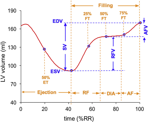
Background: Although analysis of cardiac magnetic resonance (CMR) images provides accurate and reproducible measurements of left ventricular (LV) volumes, these measurements are usually not performed throughout the cardiac cycle because of lack of tools that would allow such analysis within a reasonable timeframe. A fully-automated machine-learning (ML) algorithm was recently developed to automatically generate LV volume-time curves. Our aim was to validate ejection and filling parameters calculated from these curves using conventional analysis as a reference.
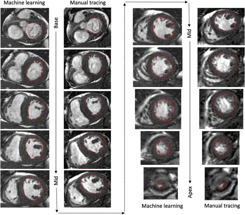
Methods: We studied 21 patients undergoing clinical CMR examinations. LV volume-time curves were obtained using the ML-based algorithm (Neosoft), and independently using slice-by-slice, frame-by-frame manual tracing of the endocardial boundaries. Ejection and filling parameters derived from these curves were compared between the two techniques. For each parameter, Bland-Altman bias and limits of agreement (LOA) were expressed in percent of the mean measured value.
Results: Time-volume curves were generated using the automated ML analysis within 2.5 ± 0.5 min, considerably faster than the manual analysis (43 ± 14 min per patient, including ~10 slices with 25-32 frames per slice). Time-volume curves were similar between the two techniques in magnitude and shape. Size and function parameters extracted from these curves showed no significant inter-technique differences, reflected by high correlations, small biases (<10%) and mostly reasonably narrow LOA.
Conclusion: ML software for dynamic LV volume measurement allows fast and accurate, fully automated analysis of ejection and filling parameters, compared to manual tracing based analysis. The ability to quickly evaluate time-volume curves is important for a more comprehensive evaluation of the patient's cardiac function.
Keywords: Artificial intelligence; Left ventricle; Time-volume curves.
Copyright © 2019 Elsevier Inc. All rights reserved.
Conflict of interest statement
Declaration of competing interest ARP has received research support from Philips Healthcare and Neosoft. Akhil Narang was funded by a T32 Cardiovascular Sciences Training Grant (5T32HL7381) from the National Institutes of Health (USA).
Figures

References
-
- Afshin M, Ben Ayed I, Islam A, Goela A, Peters TM, Li S. Global assessment of cardiac function using image statistics in MRI. Med Image Comput Comput Assist Interv 2012;15:535–43. - PubMed
-
- Tan LK, Liew YM, Lim E, McLaughlin RA. Convolutional neural network regression for short-axis left ventricle segmentation in cardiac cine MR sequences. Med Image Anal 2017;39:78–86. - PubMed
-
- Mantilla J, Paredes J, Bellanger JJ, Donal E, Leclercq C, Medina R, et al. Classification of LV wall motion in cardiac MRI using kernel Dictionary Learning with a parametric approach. Conf Proc IEEE Eng Med Biol Soc 2015;2015:7292–5. - PubMed
-
- Emad O, Yassine IA, Fahmy AS. Automatic localization of the left ventricle in cardiac MRI images using deep learning. Conf Proc IEEE Eng Med Biol Soc 2015;2015:683–6. - PubMed
Publication types
MeSH terms
Grants and funding
LinkOut - more resources
Full Text Sources
Medical
