

Methods to measure, model and manipulate fluid flow in brain
- PMID: 31838183
- PMCID: PMC7607555
- DOI: 10.1016/j.jneumeth.2019.108541
Methods to measure, model and manipulate fluid flow in brain
Abstract
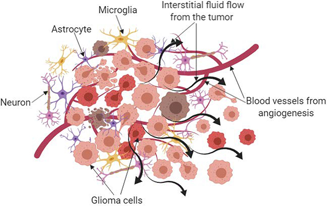
The brain consists of a complex network of cells and matrix that is cushioned and nourished by multiple types of fluids: cerebrospinal fluid, blood, and interstitial fluid. The movement of these fluids through the tissues has recently gained more attention due to implications in Alzheimer's Disease and glioblastoma. Therefore, methods to study these fluid flows are necessary and timely for the current study of neuroscience. Imaging modalities such as magnetic resonance imaging have been used clinically and pre-clinically to image flows in healthy and diseased brains. These measurements have been used to both parameterize and validate models of fluid flow both computational and in vitro. Both of these models can elucidate the changes to fluid flow that occur during disease and can assist in linking the compartments of fluid flow with one another, a difficult challenge experimentally. In vitro models, though in limited use with fluid flow, allow the examination of cellular responses to physiological flow. To determine causation, in vivo methods have been developed to manipulate flow, including both physical and pharmacological manipulations, at each point of fluid movement of origination resulting in exciting findings in the preclinical setting. With new targets, such as the brain-draining lymphatics and glymphatic system, fluid flow and tissue drainage within the brain is an exciting and growing research area. In this review, we discuss the methods that currently exist to examine and test hypotheses related to fluid flow in the brain as we attempt to determine its impact on neural function.
Keywords: Cerebrospinal fluid; Computational modeling; Glymphatic; In vitro models; Interstitial flow; Lymphatics; MRI.
Copyright © 2019. Published by Elsevier B.V.
Figures


References
-
- Arbel-Ornath M, Hudry E, Eikermann-Haerter K, Hou S, Gregory JL, Zhao L, Betensky RA, Frosch MP, Greenberg SM, Bacskai BJ, 2013a. Interstitial fluid drainage is impaired in ischemic stroke and Alzheimer’s disease mouse models. Acta Neuropathol 126, 353–364. 10.1007/s00401-013-1145-2 - DOI - PMC - PubMed
Publication types
MeSH terms
Grants and funding
LinkOut - more resources
Full Text Sources
